Review
. 2016 Jul;22(7):545-564.
doi: 10.1016/j.molmed.2016.05.003. Epub 2016 Jun 3.

Autoinflammatory Skin Disorders: The Inflammasomme in Focus

Affiliations
Review

Autoinflammatory Skin Disorders: The Inflammasomme in Focus

Prajwal Gurung et al. Trends Mol Med. 2016 Jul.

Abstract

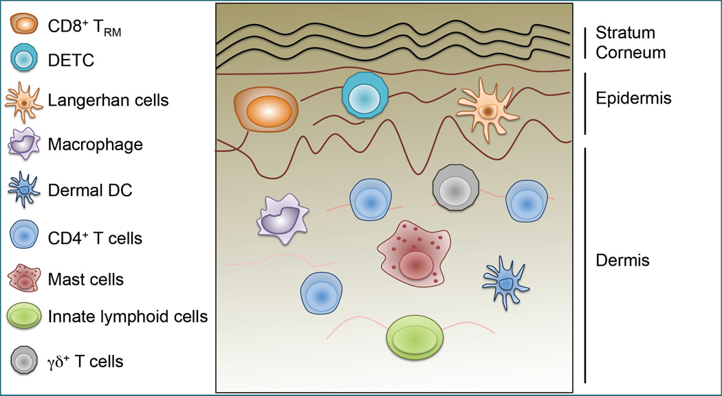
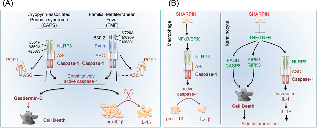
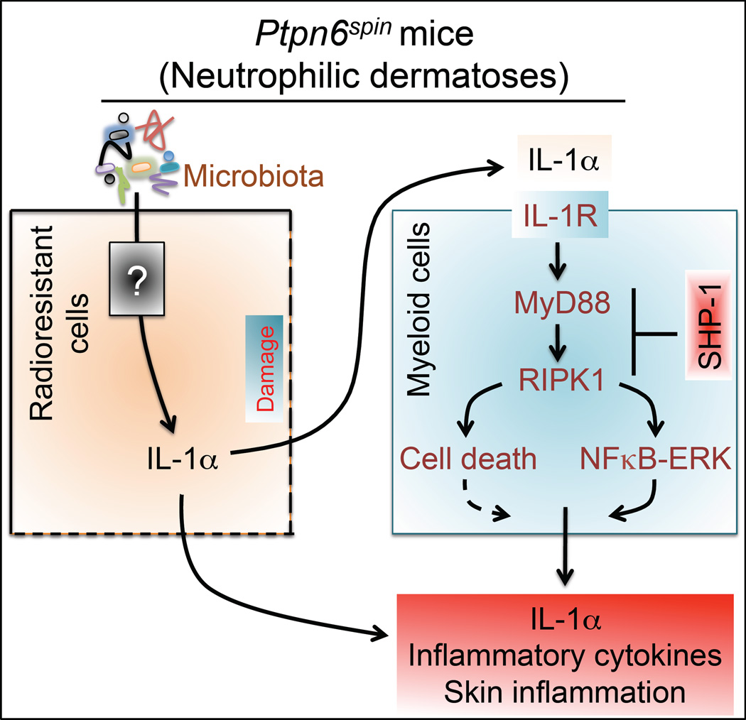
Autoinflammatory skin disorders are a group of heterogeneous diseases that include diseases such as cryopyrin-associated periodic syndrome (CAPS) and familial Mediterranean fever (FMF). Therapeutic strategies targeting IL-1 cytokines have proved helpful in ameliorating some of these diseases. While inflammasomes are the major regulators of IL-1 cytokines, inflammasome-independent complexes can also process IL-1 cytokines. Herein, we focus on recent advances in our understanding of how IL-1 cytokines, stemming from inflammasome-dependent and -independent pathways are involved in the regulation of skin conditions. Importantly, we discuss several mouse models of skin inflammation generated to help elucidate the basic cellular and molecular effects and modulation of IL-1 in the skin. Such models offer perspectives on how these signaling pathways could be targeted to improve therapeutic approaches in the treatment of these rare and debilitating inflammatory skin disorders.

Keywords: CAPS; FMF; IL-1; NLRP3; Ptpn6; Sharpin; autoimmune; inflammasome; inflammatory.


Figures

Figure 1
Figure 1. Graphical Representation of the Skin and Immune Cells Present in the Skin
The skin consists of two major layers, the epidermis and the dermis (see Box 1 and 2). The stratum corneum is an extension of the epidermal layer and consists of dead epidermal cells that provide the first major barrier for invading pathogens. The epidermis consists of mostly epidermal cells known as keratinocytes at different stages, with undifferentiated keratinocytes and melanocytes at the bottom of the epidermis, and more mature keratinocytes on the top. Major immune cell populations present in the epidemis are Langerhans cells (LC), skin resident memory CD8+ T cells (CD8+ TRM) and dendritic epidermal T cells (DETC). The dermis extends beneath the epidermis. It is composed of connective tissue and fibroblasts and is abundantly supplied with blood and lymph vessels as well as nerve endings. Immune cells that are present in the dermis include dermal dendritic cells, macrophages, T cells (CD4+ TRM and circulating CD4+), mast cells, γδ+ T cells and innate lymphoid cells (ILC).
Figure 2
Figure 2. Inflammasome Dependent and Independent Pathways Regulating IL-1 Cytokines
(A) Inflammasome promotes IL-1 and cell death: The inflammasome sensor is a cytoplasmic pathogen recongnition receptor (PRR) which senses pathogen- and damage-associated molecular patterns (DAMPs and PAMPs, respectively). When an inflammasome sensor recognizes its ligand, it recruits an adaptor, apoptosis associated speck containing a capase activation and recruitment domain (ASC). ASC further recruits caspase-1 to the complex forming the inflammasome. Within the inflammasome, autocleavage of caspase-1 results in its activation. Active caspase-1 then processes pro-IL-1β into bioactive IL-1β and activates gasdermin-D which executes a form of cell death, termed pyroptosis (Box 3). (B) Inflammasome-independent pathways that regulate IL-1β production: Activation of cell death pathways through CD95, or drug-induced mitochondrial and endoplasmic reticulum damage are known to activate caspase-8. Similarly, fungal infection can signal through the Dectin-Syk pathway to induce formation of a non-caonical complex that also activates caspase-8. Active caspase-8 processes pro-IL-1β into its mature form and promotes inflammation. In addition to caspase-8, certain proteases such as cathepsins, proteinase-3, elastase, granzyme-A, chymase, matrix metalloproteinase 9 and chymotrypsin are also known to cleave pro-IL-1β.
Figure 3
Figure 3. Mouse Models of Autoinflammatory Skin Diseases Triggered by Inflammasomes
(A) Cryopyrin-associated periodic syndrome (CAPS): Mouse models of CAPS were generated by mutating murine Nlrp3 with the same gain-of-function mutations (R258W, A350V and L351P) reported in humans. Unlike wild type (WT) NLRP3, these mutant NLRP3 form spontaneous inflammasome complexes in response to TLR priming signals and produce enhanced levels of IL-1β cytokines. Mice expressing these point mutations develop skin inflammation and disease similar to CAPS in humans. Overexpression of Pyrin domain-only protein 1 (POP1) sequesters ASC and inhibits aberrant inflammasome activation and IL-1β production. Familial-Mediterrenean fever (FMF): Mice expressing a chimeric PYRIN protein containing a mutant form of the human B30.2 domain (M680I, M690V and V726A mutations) model FMF in humans. These chimeric PYRIN expressing mice develop spontaneuos autoinflammation and skin lesions mediated by assembly of the PYRIN inflammasome. Based on the ability of POP1 to sequester ASC, we propose that POP1 overexpression can also reverse FMF disease in these mice. (B) Chronic proliferative dermatitis mutation (cpdm) in Sharpin mice (Sharpincpdm) results in SHARPIN-deficiency, which leads to spontaneous inflammmation and severe dermatitis. SHARPIN is centrally required in macrophages for NFκB and ERK signaling and promoting NLRP3 inflammasome activation. In contrast, SHARPIN-deficiency promotes NLRP3 inflammasome activation and IL-1β production in inflamed skin (keratinocytes). SHARPIN also inhibits aberrant cell death downstream of TNF-signaling (FADD-caspase-8 promotes apoptosis, RIPK1-RIPK3 promotes necroptosis) to control skin inflammation in keratinocytes.
Figure 4
Figure 4. Ptpn6spin Mice Model Human Neutrophilic Dermatosis
The microbiota may regulate IL-1α protein expression in radioresistant cells (keratinocytes) by an unclear mechanism. IL-1α released from keratinocytes during microabrasion or skin damage can recruit myeloid cells, which respond to IL-1α by producing more IL-1α and other inflammatory cytokines. Enhanced cytokine production by myeloid cells depends on MyD88-RIPK1 and NFκB-ERK signaling pathways, which are negatively regualted by SHP-1 (protein encoded by Ptpn6). Thus, SHP-1 prevents aberrant cytokine production to inhibit skin inflammation and neutrophilic dermatosis.

References

    1. Pasparakis M, et al. Mechanisms regulating skin immunity and inflammation. Nat Rev Immunol. 2014;14:289–301. - PubMed
    1. Grice EA, et al. Topographical and temporal diversity of the human skin microbiome. Science. 2009;324:1190–1192. - PMC - PubMed
    1. Gallo RL, Nakatsuji T. Microbial symbiosis with the innate immune defense system of the skin. J Invest Dermatol. 2011;131:1974–1980. - PMC - PubMed
    1. Iwase T, et al. Staphylococcus epidermidis Esp inhibits Staphylococcus aureus biofilm formation and nasal colonization. Nature. 2010;465:346–349. - PubMed
    1. Naik S, et al. Commensal-dendritic-cell interaction specifies a unique protective skin immune signature. Nature. 2015;520:104–108. - PMC - PubMed

Publication types

MeSH terms