. 2016 Dec 15;22(24):6204-6216.
doi: 10.1158/1078-0432.CCR-15-3059. Epub 2016 Jun 7.

Targeting Estrogen Receptor Signaling with Fulvestrant Enhances Immune and Chemotherapy-Mediated Cytotoxicity of Human Lung Cancer

Affiliations

Targeting Estrogen Receptor Signaling with Fulvestrant Enhances Immune and Chemotherapy-Mediated Cytotoxicity of Human Lung Cancer

Duane H Hamilton et al. Clin Cancer Res. .

Abstract

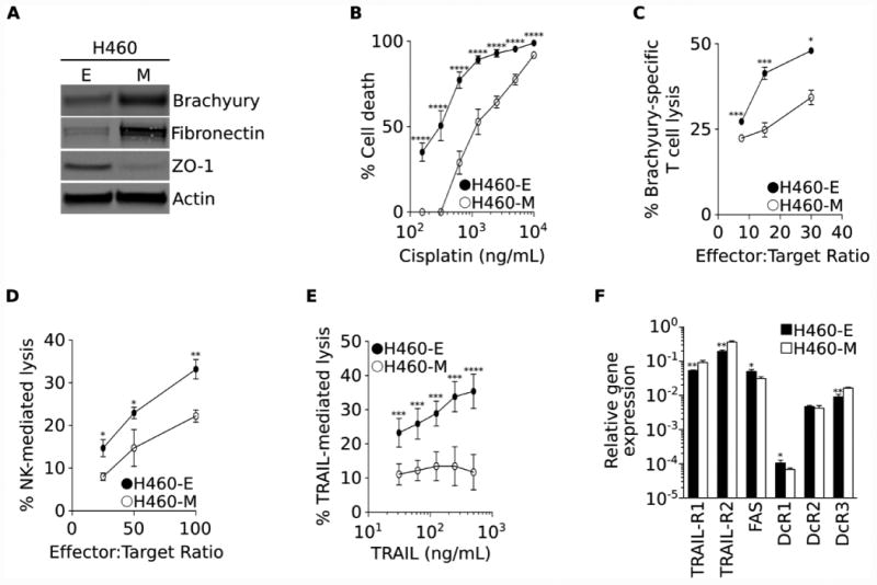
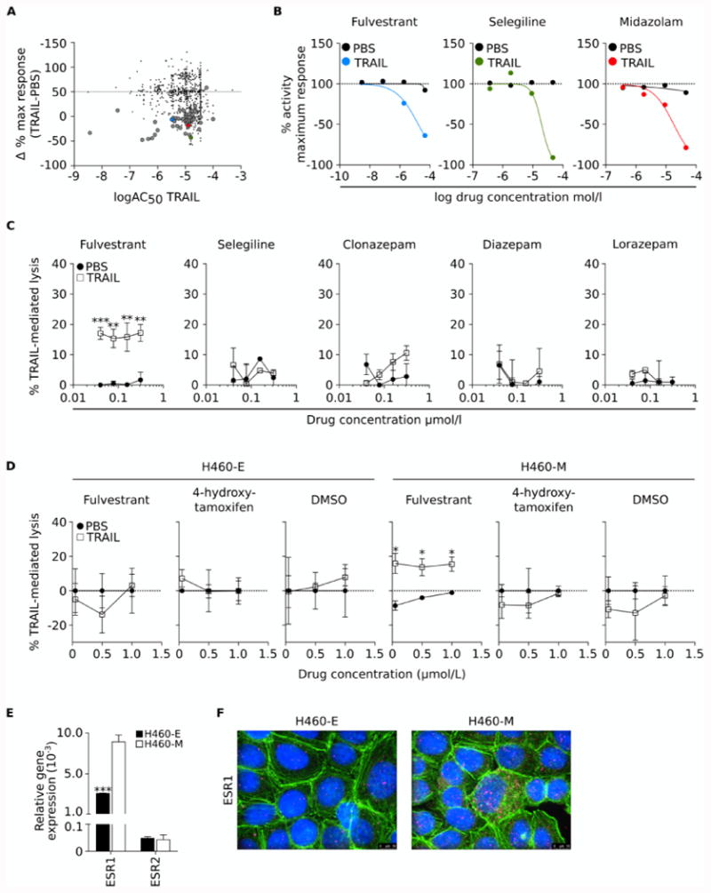
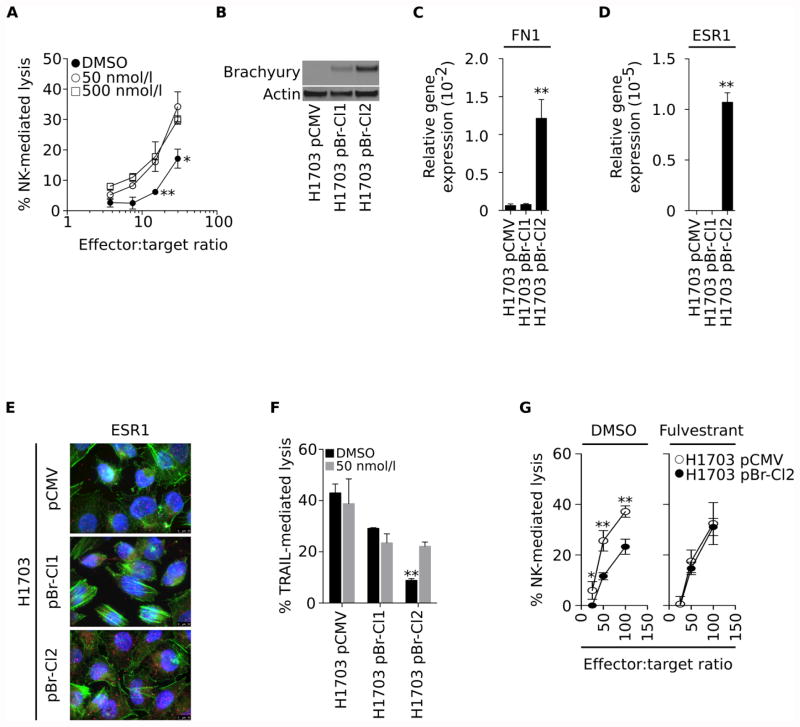
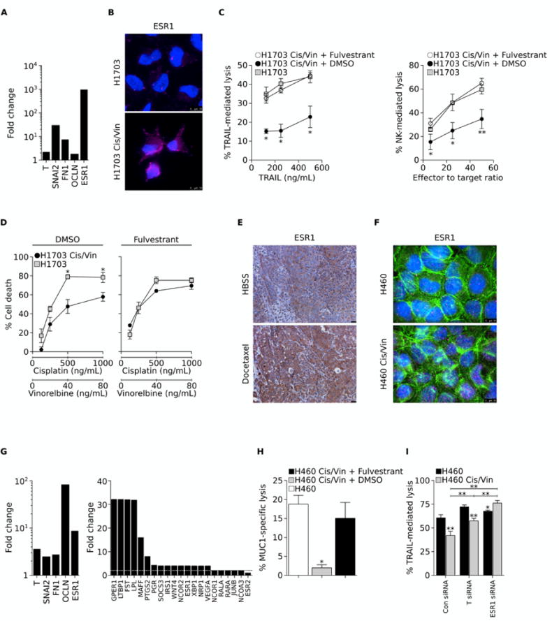
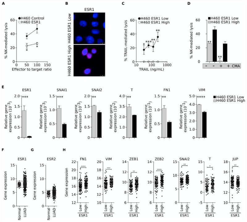
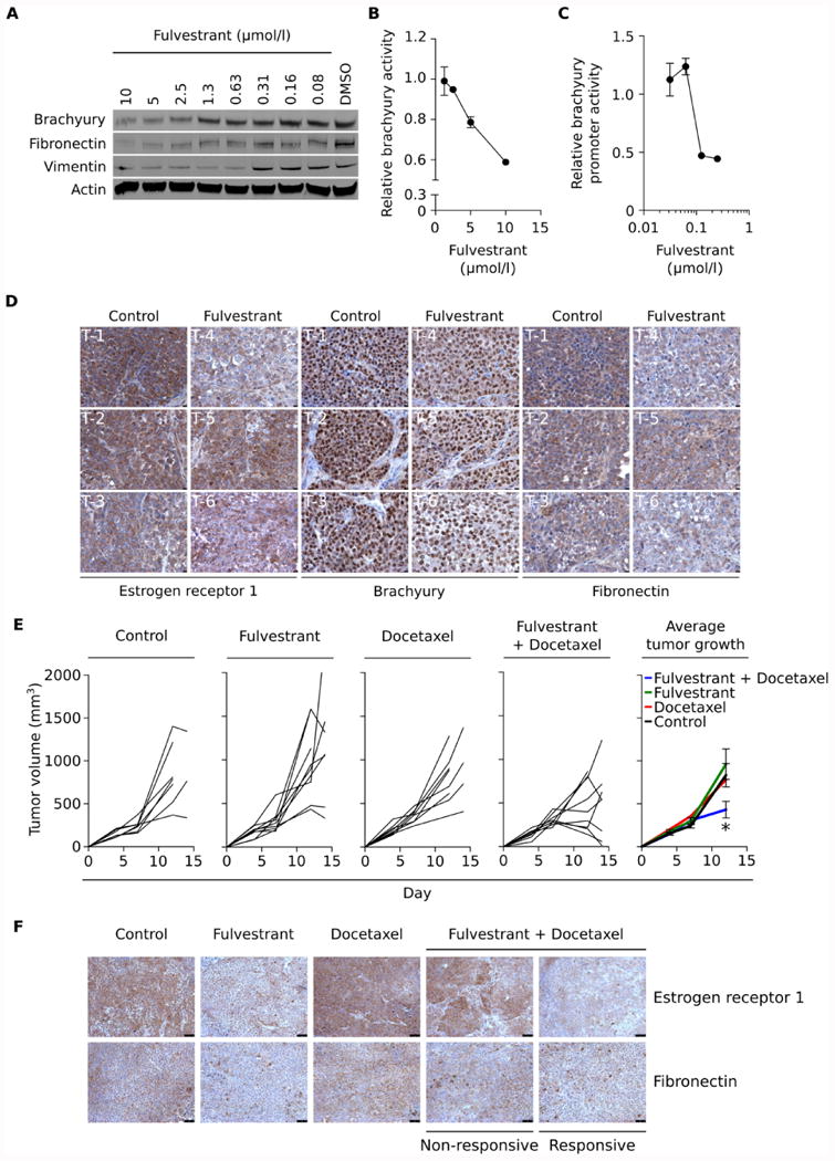
Purpose: The conversion of tumor cells from an epithelial to a mesenchymal-like phenotype, via a process designated as the epithelial-mesenchymal transition (EMT), is known to mediate tumor resistance to a variety of cell death inducers, including cytotoxic effector immune cells. The goal of this study was to identify and potentially repurpose FDA-approved compounds capable of reducing mesenchymal features of human lung carcinoma cells, which could be used in combination with immunotherapies or chemotherapeutic strategies to improve clinical responses.

Experimental design: In the current report, we have utilized a quantitative high-throughput screening (qHTS) of a pharmaceutical collection of more than 2,000 compounds to identify clinically approved drugs capable of augmenting the sensitivity of mesenchymal-like, lung cancer cells to immune- and chemotherapy-mediated lysis, both in vitro and in vivo RESULTS: The estrogen receptor antagonist fulvestrant was shown to reduce mesenchymal features of lung carcinoma cells, resulting in tumor sensitization to the cytotoxic effect of antigen-specific T cells, natural killer (NK) effector cells, and chemotherapy both in vivo and in vitro CONCLUSIONS: To our knowledge, this is the first report defining a potential role for estrogenic signaling in promoting tumor resistance to immune-mediated cytotoxicity and chemotherapy in lung cancer. Our data demonstrate a robust association between the acquisition of mesenchymal attributes, therapeutic resistance of lung carcinoma cells, and the expression of estrogen receptor 1 (ESR1), supporting further investigations on the role of estrogen signaling in lung cancer progression via the induction of EMT. Clin Cancer Res; 22(24); 6204-16. ©2016 AACR.


Conflict of interest statement

The authors have no conflict of interest to declare.

Figures

Figure 1
Figure 1. H460-M clone exhibits resistance to cell death
(A) Western blot analysis of indicated proteins expressed by the H460-E and H460-M clones. (B) Susceptibility of H460-E vs. -M clones to a range of indicated concentrations of cisplatin. (C) Percent of lysis mediated by brachyury-specific CD8+ T cells, (D) NK effector cells, and (E) recombinant TRAIL. (F) Expression of mRNA encoding for TRAIL, FAS, and decoy receptors (DcR1, DcR2, DcR3), relative to GAPDH. Error bars indicate the standard error of the mean (SEM) of triplicate measurements. [* p<0.05, ** p<0.01, *** p<0.001, **** p<0.0001].
Figure 2
Figure 2. Fulvestrant renders mesenchymal cells more sensitive to immune-mediated lysis
(A) Graphical depiction of compounds having measurable activity in the qHTS assay. The y-axis represents the Δ%MaxResponse= [(MaxResponseTRAIL+compound)-(MaxResponsePBS+compound)], where Maximum Response is the % activity at the maximum concentration of compound tested (46 μmol/L). The x-axis represents the logAC50 for TRAIL, calculated as indicated in the Materials and Methods section. Large circles represent 53 hits with curves class of 4 when used with PBS, and curves classes -1 or -2 when used with TRAIL. The top three ranked compounds are indicated by color circles: fulvestrant (blue), selegiline (green) and midazolam (red). (B) Maximum response (%) of the top three ranked compounds when used in combination with TRAIL vs. PBS. (C) Secondary screening of top compounds showing dose response curves when used in combination with TRAIL vs. PBS in H460 cells. (D) Dose response curves of H460-E and H460-M cells treated with indicated doses of fulvestrant, 4-hydroxytamoxifen (active metabolite of the FDA-approved estrogen receptor antagonist tamoxifen) or DMSO to TRAIL-mediated lysis. (E) ESR1 and ESR2 mRNA expression levels in the H460-E and -M clones, relative to GAPDH. (F) Immunofluorescent analysis of ESR1 (pink signal) expression in the H460-E and -M clones (100× magnification). Green signal corresponds to phalloidin staining; blue corresponds to DAPI. [* p<0.05, ** p<0.01, *** p<0.001].
Figure 3
Figure 3. Fulvestrant renders mesenchymal cells more sensitive to immune-mediated lysis
(A) Susceptibility of parental H460 cells treated with two doses of fulvestrant vs. DMSO to lysis by NK cells at various effector-to-target ratios. (B) Brachyury protein levels and expression of mRNA encoding for fibronectin (C) and ESR1 (D), relative to GAPDH, in clonally-derived H1703 cells transfected with pCMV vs. pBr (Clones 1 and 2). (E) Immunofluorescent analysis of ESR1 (pink signal) protein expression in H1703 pCMV, pBr-Cl1 and pBr-Cl2 cells (100× magnification). Green and blue correspond to phalloidin and DAPI staining, respectively. (F) Susceptibility to TRAIL-mediated lysis in cells pre-treated with fulvestrant (grey bars) vs. DMSO (black bars). (G). Susceptibility of H1703 pCMV and H1703 pBr-Cl2 cells treated with fulvestrant (right panel) vs. DMSO (left panel) to lysis by NK cells at various effector-to-target ratios. Error bars indicate the standard error of the mean (SEM) of triplicate measurements. [* p<0.05, ** p<0.01].
Figure 4
Figure 4. Fulvestrant reverts immune resistance of chemo-resistant lung cancer cells
(A) Fold change in expression levels of indicated mRNA in chemo-resistant vs. control H1703 cells. (B) Immunofluorescent analysis of ESR1 (pink signal) in control and cisplatin/vinorelbine-resistant (Cis/Vin) H1703 cells. Blue signal corresponds to DAPI staining. (C) Susceptibility of control H1703 vs. Cis/Vin-resistant H1703 cells to lysis by either TRAIL (left panel) or NK cells (right panel). Chemo-resistant cells were treated with DMSO or fulvestrant for 72 hours prior to the cytotoxic assay. (D) Sensitivity of control vs. Cis/Vin-resistant H1703 cells to a combination of indicated concentrations of vinorelbine and cisplatin; tumor cells were treated with DMSO (left panel) or fulvestrant (right panel) for 72 hours prior to exposure to chemotherapy. (E) Immunohistochemical analysis of ESR1 expression in H460 xenografts from mice treated with either HBSS or docetaxel (20× magnification). (F) Immunofluorescent analysis of ESR1 expression (pink signal) in control and Cis/Vin-resistant H460 cells (100× magnification). Green and blue correspond to phalloidin and DAPI staining, respectively. (G) Fold-change in expression levels of indicated mRNA in Cis/Vin-resistant vs. control H460 cells (H) Susceptibility of parental vs. chemo-resistant H460 cells to lysis by MUC1-specific CD8+ T cells. Chemo-resistant cells were treated with DMSO or fulvestrant for 72 hours prior to the cytotoxic assay. (I) TRAIL-mediated lysis of parental H460 vs. chemo-resistant H460 cells previously transfected with a control non-targeting Con siRNA vs. a pool of siRNAs targeting either brachyury (T siRNA) or the estrogen receptor 1 (ESR1 siRNA). Error bars indicate the standard error of the mean (SEM) of triplicate measurements. [* p<0.05, ** p<0.01].
Figure 5
Figure 5. Estrogen receptor mediates resistance to immune attack
(A) H460 cells stably transfected with pCMV or a vector encoding the ESR1 gene were assessed for their sensitivity to NK-mediated lysis. (B) Immunofluorescent analysis of ESR1 expression (pink signal) in single cell clones of H460 cells with High vs. Low ESR1 expression. Blue signal corresponds to DAPI staining. (C) Clones were evaluated for their susceptibility to lysis by TRAIL (C) or NK cells that were either untreated or pre-treated with CMA to inhibit perforin-dependent lytic pathways (D). (E) Relative expression of indicated mRNA in clonal H460 ESR1-High vs. ESR1-Low cells. (F) ESR1 and (G) ESR2 mRNA in normal lung vs. lung adenocarcinoma (LUAD) tissues. Shaded areas correspond to the normal range of expression for each gene, calculated as the mean expression in normal lung tissues (± two standard deviations). (H) mRNA expression of indicated genes in lung samples categorized as either ESR1 Low or High, based on the expression in normal lung tissues. Error bars indicate the standard deviation of the mean. [* p<0.05, ** p<0.01, *** p<0.001, **** p<0.0001].
Figure 6
Figure 6. Fulvestrant treatment reduces EMT markers and increases sensitivity of lung xenografts to docetaxel
(A) Western blot analysis of brachyury, fibronectin and vimentin protein in H460 cells treated in culture for six days with indicated concentrations of fulvestrant vs DMSO. (B) Brachyury transcriptional activity in H460 cells stably transfected with a brachyury-reporter vector treated for 48 hours with indicated doses of fulvestrant and normalized to DMSO-treated cells. (C) Brachyury promoter activity in H460 cells transiently transfected with a brachyury promoter vs. a GAPDH promoter reporter vector, after treatment for six days with indicated concentrations of fulvestrant. (D) Estrogen receptor 1, brachyury and fibronectin expression in H460 tumor xenografts five days after a single injection of either HBSS or fulvestrant. (E) Tumor volume of H460 xenografts treated as indicated, with fulvestrant (250 mg/kg) given on days 4 and 11 and docetaxel (20 mg/kg) on days 7 and 10. (F) Estrogen receptor 1 and fibronectin expression in H460 tumor xenografts, treated as indicated. Error bars indicate the standard error of the mean (SEM) of triplicate measurements. [* p<0.05, ** p<0.01].

References

    1. Thiery JP, Acloque H, Huang RY, Nieto MA. Epithelial-mesenchymal transitions in development and disease. Cell. 2009;139:871–90. - PubMed
    1. Kalluri R, Weinberg RA. The basics of epithelial-mesenchymal transition. J Clin Invest. 2009;119:1420–8. - PMC - PubMed
    1. Polyak K, Weinberg RA. Transitions between epithelial and mesenchymal states: acquisition of malignant and stem cell traits. Nature reviews Cancer. 2009;9:265–73. - PubMed
    1. Huang B, Cohen JR, Fernando RI, Hamilton DH, Litzinger MT, Hodge JW, et al. The embryonic transcription factor Brachyury blocks cell cycle progression and mediates tumor resistance to conventional antitumor therapies. Cell death & disease. 2013;4:e682. - PMC - PubMed
    1. Mitra A, Mishra L, Li S. EMT, CTCs and CSCs in tumor relapse and drug-resistance. Oncotarget. 2015;6:10697–711. - PMC - PubMed

MeSH terms