Review
. 2016 Sep;40(5):774-94.
doi: 10.1093/femsre/fuw009. Epub 2016 Jun 5.

Chemical probes of quorum sensing: from compound development to biological discovery

Affiliations
Review

Chemical probes of quorum sensing: from compound development to biological discovery

Michael A Welsh et al. FEMS Microbiol Rev. 2016 Sep.

Abstract

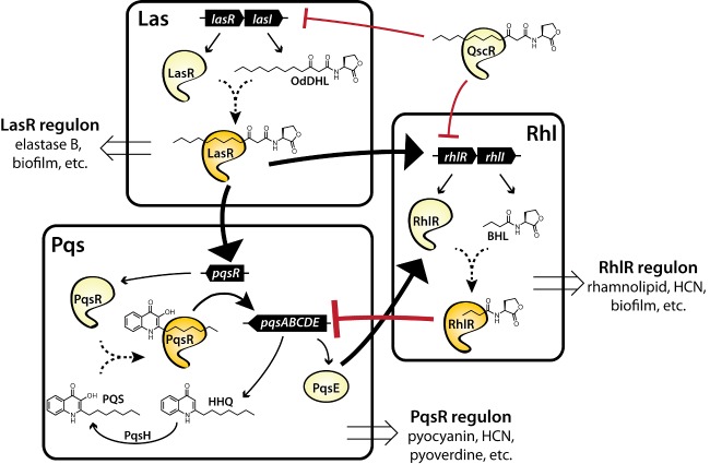
Bacteria can utilize chemical signals to coordinate the expression of group-beneficial behaviors in a method of cell-cell communication called quorum sensing (QS). The discovery that QS controls the production of virulence factors and biofilm formation in many common pathogens has driven an explosion of research aimed at both deepening our fundamental understanding of these regulatory networks and developing chemical agents that can attenuate QS signaling. The inherently chemical nature of QS makes studying these pathways with small molecule tools a complementary approach to traditional microbiology techniques. Indeed, chemical tools are beginning to yield new insights into QS regulation and provide novel strategies to inhibit QS. Here, we review the most recent advances in the development of chemical probes of QS systems in Gram-negative bacteria, with an emphasis on the opportunistic pathogen Pseudomonas aeruginosa We first describe reports of novel small molecule modulators of QS receptors and QS signal synthases. Next, in several case studies, we showcase how chemical tools have been deployed to reveal new knowledge of QS biology and outline lessons for how researchers might best target QS to combat bacterial virulence. To close, we detail the outstanding challenges in the field and suggest strategies to overcome these issues.

Keywords: LuxR-type receptor; Pseudomonas aeruginosa; acylated homoserine lactone; antivirulence; chemical biology; quorum sensing.


Figures

Graphical Abstract Figure.
We describe the development and application of novel chemical tools to interrogate bacterial quorum sensing pathways—with a particular focus on the Gram-negative pathogen Pseudomonas aeruginosa—and outline several future challenges for this approach.
Figure 1.
Figure 1.
The canonical LuxI-LuxR QS circuit from V. fischeri. The LuxI synthase produces an AHL autoinducer that accumulates in the surroundings at a concentration proportional to cell density. At a threshold level, productive binding between the AHL and a cognate LuxR receptor occurs resulting in receptor dimerization and association with QS-responsive promoters. Figure adapted with permission from Moore et al. (2015). Copyright 2015 American Chemical Society.
Figure 2.
Figure 2.
The QS system in P. aeruginosa. Once activated by its native ligand, OdDHL, LasR is able to induce expression and activation of the Rhl and Pqs systems. RhlR suppresses Pqs signaling by inhibiting expression of the pqsA-E operon. Pqs augments the Rhl system through an unknown mechanism, possibly involving PqsE. In turn, Las and Rhl are repressed by the ‘orphan’ LuxR-type receptor QscR. The three main QS systems work in tandem to control the global expression of virulence phenotypes (exoenzyme production, swarming, biofilm formation, etc.) with Las, Rhl and Pqs each being primarily associated with elastase B, rhamnolipid and pyocyanin production, respectively. Large arrows indicate major regulatory relationships between circuits. Solid arrowheads indicate positive regulation, while flat, red arrowheads indicate negative regulation. Figure adapted with permission from Welsh et al. (2015). Copyright 2015 American Chemical Society.
Figure 3.
Figure 3.
Examples of small molecule inhibitors and activators of LuxR-type receptors. Modulators of LasR, CviR and QscR are shown. The EC50 and IC50 values listed were determined using E. coli reporter strains, except where noted. * = IC50 value determined in a P. aeruginosa reporter strain. Compound references: 1, Amara et al. (2009); 2, Geske, Mattmann and Blackwell (2008); 3, Swem et al. (2009); 4, Mattmann et al. (2011), 5, Müh et al. (2006b); 6, Hentzer et al. (2002); 7, Müh et al. (2006a); 8, Müh et al. (2006a); Moore et al. (2015).
Figure 4.
Figure 4.
Examples of compound dose-response behavior in reporter gene assays. Agonism curves (blue) are generated by adding increasing amounts of small molecules and assaying for reporter turn-on. Antagonism curves (red) are generated by stimulating the reporter with a set concentration of native ligand and adding increasing amounts of non-native ligand. Figure adapted with permission from Moore et al. (2015). Copyright 2015 American Chemical Society.
Figure 5.
Figure 5.
Novel compounds designed to mimic native AHLs. (A) AHL mimics with non-homoserine lactone head groups that have been evaluated in several LuxR-type receptors (LasR, TraR and LuxR). (B) Selected AHL-type agonists and antagonists of the RhlR receptor from P. aeruginosa.
Figure 6.
Figure 6.
Antagonists of LasR derived from the TP scaffold. Appending a homoserine lactone or nitrophenyl head group (derived from the TP scaffold) with an aryl bromide tail mode switches potent LasR agonists to antagonists. Figure adapted with permission from O'Reilly and Blackwell (2015). Copyright 2016 American Chemical Society.
Figure 7.
Figure 7.
Small molecule modulators of LuxR-type receptors identified from natural sources or in silico screens. Compounds 17, 18, 19 and 21 are antagonists of LasR. Compound 22 is an agonist of LasR. Honaucin A, 20, is an antagonist of LuxR.
Figure 8.
Figure 8.
Irreversible inhibitors of LasR. Introduction of electrophilic groups (highlighted in green) into scaffolds known to target LasR resulted in irreversible LasR inhibition. Perez and coworkers discovered the TP analog 22 (O'Brien et al. 2015), while Meijler and coworkers developed the OdDHL mimic 23 (Amara et al. 2016).
Figure 9.
Figure 9.
Inhibitors of LuxI synthases. (A) Structures of known lead inhibitors of LuxI AHL synthases. (B) Outline of an assay developed by Greenberg and coworkers that allows high-throughput screening for inhibitors of the BmaI1 synthase. Definitions: Nuh, nucleoside hydrolase; Ade, adenine deaminase; XO, xanthine oxidase; HRP, horseradish peroxidase. Panel B is adapted with permission from Christensen et al. (2013).
Figure 10.
Figure 10.
PQS biosynthesis and small molecule inhibitors of PqsR. (A) Schematic of the biosynthesis of PQS by the enzymes PqsABCDE in P. aeruginosa. (B) Compounds prepared by Hartmann and coworkers as direct mimics of PQS. (C) Small molecule inhibitors of PqsR identified in fragment-based and high-throughput screens by Hartmann and coworkers (31, 32) and Rahme and coworkers (33).
Figure 11.
Figure 11.
Inhibitors of PqsD identified by Hartmann and coworkers. (A) Compounds 34 and 35 were designed as PqsD transition-state inhibitors. (B) Compound 36 was identified as a PqsD inhibitor, and its analog, 37, is a covalent PqsD inhibitor. (C) Discovery of a dual PqsD/PqsR inhibitor, 38, by structural reduction from previously identified compounds.
Figure 12.
Figure 12.
Barriers to the spread of resistance to quorum sensing inhibitors (QSIs). A schematic is shown of two possible barriers to the spread of resistance to QSIs in a bacterial population. (A) Should a bacterium become resistant to a QSI via a ‘signal-dependent’ mechanism (e.g. via receptor mutation that prevents inhibitor binding, upregulation of efflux pumps, etc.), a rare, resistant mutant in the population cannot produce enough autoinducer to activate its QS system(s). (B) If a bacterium becomes resistant via a ‘signal-independent’ mechanism (i.e. via a receptor mutation that leads to constitutive activation or converts the QSI to an agonist), the bacterium is able to produce QS-regulated virulence factors. However, these excreted ‘public goods’ can be utilized by nearby cheaters, which gain a fitness advantage. Adapted with permission from Gerdt and Blackwell (2014). Copyright 2014 American Chemical Society.
Figure 13.
Figure 13.
X-ray crystal structures of TraR bound to its native AHL, SdiA in the AHL-bound and apo forms, and CviR bound to an inhibitor (CL). (A) Structure of the [TraR:OOHL]2 complex bound to DNA. When bound to its native AHL ligand, OOHL, TraR exists as a homodimer capable of binding DNA and regulating transcription. Image adapted from Zhang et al. (2002); PDB ID 1L3L. (B) Structure of the [SdiA:OHHL]2 complex; OHHL = N-3-oxo-hexanoyl L-homoserine lactone. Image adapted from Nguyen et al. (2015); PDB ID 4Y15. (C) Structure of [SdiA]2 crystalized in the absence of AHL. Further examination of this structure revealed the presence of 1-octanoyl-rac-glycerol in the AHL-binding pocket; see the text. Image adapted from Nguyen et al. (2015); PDB ID 4Y13. (D) Structure of the [CviR:CL]2 complex. When bound to the inhibitor CL, CviR can homodimerize, but adopts a ‘crossed domain’ conformation that cannot bind DNA. For the structure of CL (3), see Fig. 3. Image adapted from Chen et al. (2011); PDB ID 3QP5.
Figure 14.
Figure 14.
Determination of residues in the LasR ligand-binding pocket responsible for receptor activation or inhibition by non-native ligands. (A) Small molecule LasR ligands of similar structure often have inverse activities. Indicated is the compound activity in WT LasR. (B) Mutation of select residues in the LasR ligand-binding pocket (e.g. W60, Y56 and S129) can invert the activity of some LasR ligands. Panel B is adapted with permission from Gerdt et al. (2014). Copyright 2014 Elsevier.
Figure 15.
Figure 15.
Fluorescence labeling of LasR in live cells using small molecule probes. (A) Once bound to LasR, the small molecule OdDHL mimic 1 can be captured with a BODIPY probe via a bioorthogonal oxime ligation. (B) Fluorescence image of P. aeruginosa cells treated with compound 1 from panel A that indicates localization of LasR at the cell poles. Pseudomonas aeruginosa cells are outlined for clarity. Figure adapted with permission from Rayo et al. (2011). Copyright 2011 American Chemical Society.
Figure 16.
Figure 16.
Small molecule LasR antagonists are substrates of active efflux pumps. (A) Plot of compound percent LasR inhibition in an E. coli reporter strain vs in a P. aeruginosa reporter strain with active efflux pumps. (B) When the efflux pump MexAB-OprM is deleted, compound activity in P. aeruginosa positively correlates with activity in E. coli. Figure adapted with permission from Moore et al. (2014). Copyright 2014 John Wiley & Sons.
Figure 17.
Figure 17.
Environmental factors affecting QS activation and small molecule activity in P. aeruginosa. (A) Schematic of a P. aeruginosa cell illustrating how external environmental factors can influence QS pathways. Iron concentrations may activate the Pqs system indirectly through the regulatory RNA PrrF (Oglesby et al. 2008). Phosphate levels are known to induce Rhl and Pqs through the PhoR-PhoB two component system (Jensen et al. 2006). Carbon catabolite repression can influence QS activity through downregulation of Lon protease (Yang et al. 2015), a post-translational regulator of Las and Rhl. The stringent response differentially activates certain QS systems though ppGpp-binding transcription factors (Schafhauser et al. 2014). (B) Example of differential modulation of a QS phenotype by a small molecule in different environmental conditions. Under phosphate-rich conditions (filled shapes), the LasR antagonist 5 strongly inhibits P. aeruginosa pyocyanin production. In phosphate-depleted conditions (open shapes), the compound is inactive. (C) Small molecule probes that have been used to investigate the environmental requirements for QS inhibition in P. aeruginosa. Compounds utilized include the LasR antagonist 5 (V-06-018) (Müh et al. ; Moore et al. 2015), the RhlR antagonist 14 (E22) (Eibergen et al. ; Welsh et al. 2015), the RhlR agonist 13 (S4) (Welsh et al. 2015) and the PqsR antagonist 33 (M64) (Starkey et al. 2014). QS receptor control of pyocyanin, rhamnolipid and elastase B production is illustrated with solid arrows indicating direct, positive regulation and dashed arrows indicating positive regulation by indirect or unknown mechanisms. For each compound, red lines with flat ends indicate receptor antagonism, while the blue arrow indicates receptor agonism. Figure adapted with permission from Welsh and Blackwell (2016). Copyright 2016 Elsevier.

Similar articles

Cited by

References

    1. Allegretta G, Weidel E, Empting M, et al. Catechol-based substrates of chalcone synthase as a scaffold for novel inhibitors of PqsD. Eur J Med Chem. 2015;90:351–9. - PubMed
    1. Allen RC, Popat R, Diggle SP, et al. Targeting virulence: can we make evolution-proof drugs? Nat Rev Microbiol. 2014;12:300–8. - PubMed
    1. Amara N, Mashiach R, Amar D, et al. Covalent inhibition of bacterial quorum sensing. J Am Chem Soc. 2009;131:10610–9. - PubMed
    1. Amara N, Gregor R, Rayo J, et al. Fine-tuning covalent inhibition of bacterial quorum sensing. ChemBioChem. 2016;17:825–35. - PubMed
    1. Balasubramanian D, Schneper L, Kumari H, et al. A dynamic and intricate regulatory network determines Pseudomonas aeruginosa virulence. Nucleic Acids Res. 2013;41:1–20. - PMC - PubMed

MeSH terms