

Aberrant methylation-mediated silencing of microRNAs contributes to HPV-induced anchorage independence
- PMID: 27270309
- PMCID: PMC5190061
- DOI: 10.18632/oncotarget.9698
Aberrant methylation-mediated silencing of microRNAs contributes to HPV-induced anchorage independence
Abstract
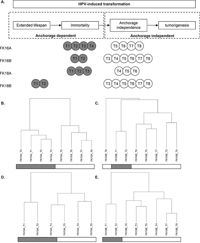
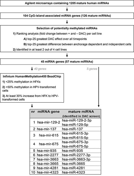
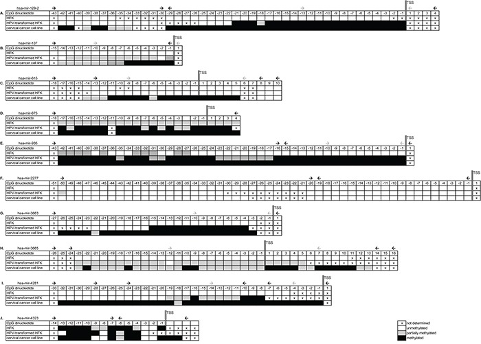
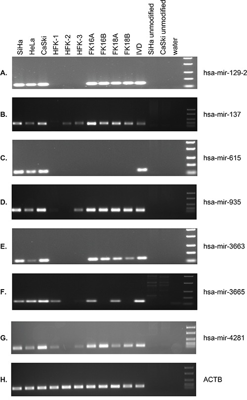
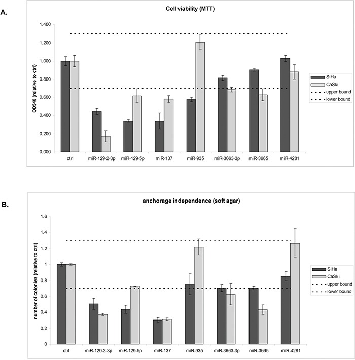
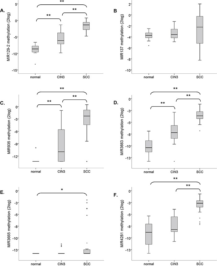
Cervical cancer and a subset of anogenital and head-and-neck carcinomas are caused by high-risk types of the human papillomavirus (hrHPV). During hrHPV-induced malignant transformation keratinocytes become able to grow anchorage independently, a tumorigenic trait at least partly associated with inactivation of tumor suppressor genes. We used hrHPV-containing keratinocytes to investigate the role of DNA methylation-mediated silencing of microRNAs (miRNAs) in the acquisition of anchorage independence.Anchorage dependent (n=11) and independent passages (n=19) of 4 hrHPV-immortalized keratinocyte cell lines were treated with 2'-deoxy-5-azacytidine (DAC). Genome-wide miRNA expression profiles before and after treatment were compared to identify miRNAs silenced by methylation. Bisulfite sequencing and methylation-specific PCR showed increased methylation of hsa-mir-129-2/-137/-935/-3663/-3665 and -4281 in anchorage independent HPV-transformed keratinocytes and cervical cancer cell lines. Mature miRNAs derived from hsa-mir-129-2/-137/-3663 and -3665 showed functional relevance as they decreased anchorage independence in cervical cancer cell lines. Cervical (pre)cancerous lesions demonstrated increased methylation of hsa-mir-129-2/-935/-3663/-3665 and -4281, underlining the clinical relevance of our findings.In conclusion, methylation-mediated silencing of tumor suppressive miRNAs contributes to acquisition of an anchorage independent phenotype. This study further substantiates the importance of miRNAs during early stages of carcinogenesis and underlines their potential as both disease markers and therapeutic targets.
Keywords: CIN lesion; DNA methylation; anoikis; cervical cancer.
Conflict of interest statement
PJFS, RDMS and CJLMM have minority stake in Self-Screen B.V., a spin-off company of VU University Medical Center Amsterdam. PJFS has been on the speaker's bureau of Roche, Abbott, Gen-Probe, Qiagen and Seegene. He is consultant for Crucell Holland B.V. CJLMM has participated in the sponsored speaker's bureau of Merck, GSK, Qiagen, Menarini, Seegene, and Roche, and served occasionally on the scientific advisory board of GSK, Qiagen, Merck, and Roche. CJLMM has occasionally been a consultant for Qiagen and Genticel and is a minority shareholder of Diassay B.V. Formerly CJLMM was a minority shareholder of Delphi Biosciences. All other authors have no conflicts of interest to declare.
Figures
Similar articles
-
Methylation-mediated transcriptional repression of microRNAs during cervical carcinogenesis.Epigenetics. 2013 Feb;8(2):220-8. doi: 10.4161/epi.23605. Epub 2013 Jan 16. Epigenetics. 2013. PMID: 23324622 Free PMC article.
-
Methylation-specific digital karyotyping of HPV16E6E7-expressing human keratinocytes identifies novel methylation events in cervical carcinogenesis.J Pathol. 2013 Sep;231(1):53-62. doi: 10.1002/path.4210. Epub 2013 Jul 8. J Pathol. 2013. PMID: 23674368
-
Focal aberrations indicate EYA2 and hsa-miR-375 as oncogene and tumor suppressor in cervical carcinogenesis.Genes Chromosomes Cancer. 2013 Jan;52(1):56-68. doi: 10.1002/gcc.22006. Epub 2012 Sep 14. Genes Chromosomes Cancer. 2013. PMID: 22987659
-
The Natural History of Cervical Cancer and the Case for MicroRNAs: Is Human Papillomavirus Infection the Whole Story?Int J Mol Sci. 2024 Dec 3;25(23):12991. doi: 10.3390/ijms252312991. Int J Mol Sci. 2024. PMID: 39684702 Free PMC article. Review.
-
[The role of microRNAs in the pathogenesis of cervical cancer and its relationship to HPV].Sheng Li Ke Xue Jin Zhan. 2012 Aug;43(4):251-6. Sheng Li Ke Xue Jin Zhan. 2012. PMID: 23189617 Review. Chinese.
Cited by
-
Epigenetic Changes Associated with Early Life Experiences: Saliva, A Biospecimen for DNA Methylation Signatures.Curr Genomics. 2018 Dec;19(8):676-698. doi: 10.2174/1389202919666180307150508. Curr Genomics. 2018. PMID: 30532647 Free PMC article.
-
Comparative Analysis of Urine Fractions for Optimal Bladder Cancer Detection Using DNA Methylation Markers.Cancers (Basel). 2020 Apr 2;12(4):859. doi: 10.3390/cancers12040859. Cancers (Basel). 2020. PMID: 32252299 Free PMC article.
-
Placental Epigenome Impacts Fetal Development: Effects of Maternal Nutrients and Gut Microbiota.Nutrients. 2024 Jun 13;16(12):1860. doi: 10.3390/nu16121860. Nutrients. 2024. PMID: 38931215 Free PMC article. Review.
-
HPV epigenetic mechanisms related to Oropharyngeal and Cervix cancers.Cancer Biol Ther. 2018;19(10):850-857. doi: 10.1080/15384047.2017.1310349. Epub 2018 May 14. Cancer Biol Ther. 2018. PMID: 28362190 Free PMC article. Review.
-
Identification of Deregulated Pathways, Key Regulators, and Novel miRNA-mRNA Interactions in HPV-Mediated Transformation.Cancers (Basel). 2020 Mar 16;12(3):700. doi: 10.3390/cancers12030700. Cancers (Basel). 2020. PMID: 32188026 Free PMC article.
References
-
- Doorbar J. Molecular biology of human papillomavirus infection and cervical cancer. Clin Sci (Lond) 2006;110:525–541. - PubMed
-
- Chen TM, Pecoraro G, Defendi V. Genetic analysis of in vitro progression of human papillomavirus-transfected human cervical cells. Cancer Res. 1993;53:1167–1171. - PubMed
-
- Freedman VH, Shin SI. Cellular tumorigenicity in nude mice: correlation with cell growth in semi-solid medium. Cell. 1974;3:355–359. - PubMed
MeSH terms
Substances
LinkOut - more resources
Full Text Sources
Other Literature Sources
Medical
Molecular Biology Databases
