. 2016 Aug;17(8):914-21.
doi: 10.1038/ni.3457. Epub 2016 Jun 6.

Pyrin inflammasome activation and RhoA signaling in the autoinflammatory diseases FMF and HIDS

Affiliations

Pyrin inflammasome activation and RhoA signaling in the autoinflammatory diseases FMF and HIDS

Yong Hwan Park et al. Nat Immunol. 2016 Aug.

Abstract

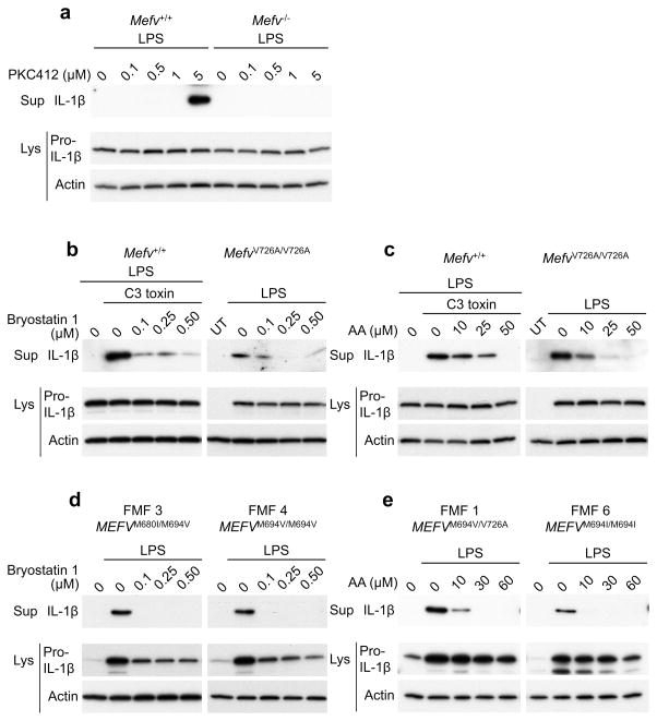
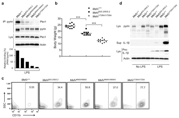
Mutations in the genes encoding pyrin and mevalonate kinase (MVK) cause distinct interleukin-1β (IL-1β)-mediated autoinflammatory diseases: familial Mediterranean fever (FMF) and hyperimmunoglobulinemia D syndrome (HIDS). Pyrin forms an inflammasome when mutant or in response to bacterial modification of the GTPase RhoA. We found that RhoA activated the serine-threonine kinases PKN1 and PKN2 that bind and phosphorylate pyrin. Phosphorylated pyrin bound to 14-3-3 proteins, regulatory proteins that in turn blocked the pyrin inflammasome. The binding of 14-3-3 and PKN proteins to FMF-associated mutant pyrin was substantially decreased, and the constitutive IL-1β release from peripheral blood mononuclear cells of patients with FMF or HIDS was attenuated by activation of PKN1 and PKN2. Defects in prenylation, seen in HIDS, led to RhoA inactivation and consequent pyrin inflammasome activation. These data suggest a previously unsuspected fundamental molecular connection between two seemingly distinct autoinflammatory disorders.


Conflict of interest statement

COMPETING FINANCIAL INTERESTS

The authors declare no competing financial interests.

Figures

Figure 1
Figure 1. The pyrin inflammasome is activated by inactivation of RhoA
(a) BMDMs from wild-type (WT) or Mefv−/− mice were primed with LPS (1 μg/ml) and C3 toxin (0.5 μg/ml) or TcdB (0.5 μg/ml) for 6h. Cell culture supernatants (Sup) and cell lysates (Lys) were analyzed by immunoblotting as indicated. (b,c) BMDMs from (b) WT mice were treated with LPS, C3 toxin, and CNF toxin, or (c) MefvV726A/V726A mice were co-treated with LPS and CNF toxin for 6h and then analyzed for IL-1β release (top) and for activated RhoA levels (bottom), % change of RhoA activity to (b) LPS or (c) UT. ** P ≤ 0.005 (unpaired two-tailed t-test). Data represent the mean ± s.e.m of n = 6 mice. (d) BMDMs from WT mice were co-treated with LPS, the indicated concentration of CNF toxin for 5h, and then ATP (2 mM) for 0.5h, flagellin (0.5 μg/ml with 25 μl/ml DOTAP) for 1h, or dsDNA (1 μg/ml with 2.5 μl/ml Lipofectamine 2000) for 1h and then analyzed for IL-1β release. (e) LPS-primed BMDMs from MefvV726A/V726A mice were treated with the indicated dose of NKH477 for 1h and then analyzed for IL-1β release (top) and for activated RhoA levels (bottom), % change of RhoA activity to LPS. ** P ≤ 0.005 (unpaired two-tailed t-test). Data represent the mean ± s.e.m of n = 8 mice. All immunoblot data shown are representative of at least three independent experiments.
Figure 2
Figure 2. Colchicine suppresses the pyrin inflammasome
(a) BMDMs from MefvV726A/V726A or WT mice were treated with the indicated concentration of colchicine without or with C3 toxin, and activated RhoA (GTP-RhoA) was assessed by using a pull-down assay with Rhotekin-RBD beads. (b) LPS-primed BMDMs from MefvV726A/V726A mice were treated with the indicated concentration of colchicine. (c) BMDMs from WT mice were co-treated with LPS, C3 toxin, and indicated concentration of colchicine. (d,f) PBMCs from (d) three FMF (designated patient number 3, 4, and 5) or (f) two CAPS (designated patient number 3 and 4) patients with the indicated mutations in MEFV or NLRP3 were co-treated with LPS and the indicated dose of colchicine for 6h. (e) BMDMs from WT mice were primed with LPS for 3h and then treated with the indicated concentration of colchicine and ATP (2 mM) for 0.5h, flagellin (0.5 μg/ml with 25 μl/ml DOTAP) for 1h, or dsDNA (1 μg/ml with 2.5 μl/ml of Lipofectamine 2000) for 1h. Cell culture supernatants and cell lysates were analyzed by immunoblotting as indicated. Immunoblot data shown are representative of one experiment (d, f) or at least three independent experiments (ac, e).
Figure 3
Figure 3. Inhibition of RhoA effector kinases activates the pyrin inflammasome
(a) LPS-primed BMDMs from WT, Casp1, Asc, Nlrp3, Nlrc4, Aim2, Mefv-deficient mice, or C-terminal pyrin-truncation mice (MefvΔCt/ΔCt) were treated with staurosporine (1 μM) for 1h. (b) BMDMs were transiently transfected with negative control (Ctrl) that has no significant sequence similarity to mouse or human gene sequences, Pkn1, Pkn2, Pkn1 + Pkn2, Rock1, Rock2, Rock1 + Rock2, or Pkn1 + Pkn2 + Rock1 + Rock2 siRNAs and then treated with LPS for 8h. (c) BMDMs from WT or Mefv−/− mice were transiently transfected with Ctrl or Pkn1 + Pkn2 siRNAs and then treated with LPS for 8h. Cell culture supernatants and cell lysates were analyzed by immunoblotting as indicated. All immunoblot data shown are representative of at least three independent experiments.
Figure 4
Figure 4. The RhoA effector kinases, PKNs, suppress pyrin inflammasome activation
(a) LPS-primed BMDMs from WT or Mefv−/− mice were treated with the indicated dose of PKC412 for 1h. (b,c) BMDMs from WT and FMF-knock-in (FMF-KI) mice were co-treated with LPS and the indicated dose of (b) bryostatin 1 or (c) arachidonic acid (AA) with or without C3 toxin for 6h. (d,e) PBMCs from FMF patients (designated patient number 1, 3, 4, and 6) with the indicated mutations in MEFV were co-treated with LPS and the indicated dose of (d) bryostatin 1 or (e) arachidonic acid for 6h. Cell culture supernatants and cell lysates were analyzed by immunoblotting as indicated. All immunoblot data shown are representative of at least three independent experiments.
Figure 5
Figure 5. PKNs bind and phosphorylate pyrin
(a) Lysates from LPS-primed BMDMs of WT and Mefv−/− mice or lysates from LPS-primed WT BMDMs co-treated with C3 toxin, C3 toxin and arachidonic acid, or C3 toxin and bryostatin 1 were immunoprecipitated with anti-pyrin antibody, and immune complexes were analyzed by immunoblot for PKN1 and pyrin. (b) The lysates of 239T cells transiently expressing human pyrin and full-length or various deleted forms of PKN1 (top) were immunoprecipitated with anti-myc (pyrin) and analyzed by immunoblot using anti-V5 (PKN1) antibody (bottom). (c) Myc&his-tagged N-terminal human pyrin (aa’s 1-330) was expressed and purified by Ni-NTA beads. Purified N-terminal pyrin was incubated with or without recombinant PKN1 or PKN2 and analyzed for phosphorylation by staining with Pro-Q Diamond reagent or immunoblotting with an antibody specific for phospho-Ser followed by Coomassie blue staining and immunoblotting with anti-myc antibody for loading control. All immunoblot and gel staining data shown are representative of at least three independent experiments.
Figure 6
Figure 6. The binding of PKN1 to the pyrin of FMF-KI mice is substantially decreased relative to wild-type mouse pyrin, which lacks a B30.2 orthologous domain, and KI mice with the WT B30.2 domain of human pyrin have a milder inflammatory phenotype than KI mice with FMF-associated mutations
(a) BMDMs from WT and FMF-KI mice with WT or the indicated FMF-associated mutant human B30.2 domain were treated with LPS. Lysates were immunoprecipitated with anti-pyrin antibody, and immune complexes were analyzed by immunoblot for PKN1 and pyrin. Densitometry analysis of PKN1 bands, normalized to the pyrin bands in immune complexes, is shown in the histogram below. (b) Body weights of 8-week-old males of Mefv+/+, MefvB30.2/B30.2, and MefvV726A/V726A mice. Each graph point represents one mouse, and means are shown as horizontal bars. *** P ≤ 0.0005 (unpaired two-tailed t-test). (c) Peripheral blood cells from 8-week-old Mefv+/+, MefvB30.2/B30.2, MefvM680I/M680I, MefvM694V/M694V, and MefvV726A/V726A male mice were analyzed for CD11b+ myeloid cells. Numbers indicate percentage of total cells in gates. Data are representative of n = 5 mice (WT and MefvM680I/M680I), n = 7 mice (MefvV726A/V726A), or n = 8 mice (MefvB30.2/B30.2 and MefvM694V/M694V). (d) Bone marrow CD11b+ cells from 8-week-old males of Mefv+/+, MefvB30.2/B30.2, and MefvV726A/V726A mice were treated with/without LPS for 6h. Cell culture supernatants and cell lysates were analyzed by immunoblotting as indicated. All immunoblot data shown are representative of at least three independent experiments.
Figure 7
Figure 7. The pyrin inflammasome is inhibited by phosphorylation and subsequent 14-3-3 binding
(a) Purified myc- and his-tagged WT N-terminal pyrin (aa’s 1-330) or N-terminal pyrin with S208A and S242A mutations was incubated with recombinant PKN1 or PKN2 and analyzed for phosphorylation by immunoblotting with an antibody specific for phospho-Ser followed by immunoblotting with anti-myc antibody. (b) WT BMDMs were treated with LPS, C3 toxin, and arachidonic acid or bryostatin 1. (c) BMDMs from WT and FMF-KI mice with WT or the indicated mutant human B30.2 domain were treated with LPS. (b, c) Lysates were immunoprecipitated with anti-mouse pyrin antibody, and immune complexes were analyzed by immunoblot for 14-3-3ε. (d) WT or the indicated FMF-associated human mutant pyrin proteins were transiently expressed in 239T cells. (e) Macrophages differentiated from PBMCs of 4 healthy controls and 5 FMF patients were treated with IFN-γ for 16h to induce pyrin expression, and primed with LPS for 6h. (d, e) The lysates were immunoprecipitated with anti-human pyrin antibody, and immune complexes were analyzed by immunoblot for 14-3-3ε. (f) BMDMs from WT and Mefv−/− mice were transfected with Ctrl or 14-3-3ε and 14-3-3τ siRNAs and treated with LPS for 8h. Cell culture supernatants and cell lysates were analyzed by immunoblotting as indicated. (g) U937 cells transiently expressing WT or the indicated mutant pyrin proteins were primed with LPS for 6h. Cell culture supernatants and cell lysates were analyzed by immunoblotting as indicated. All immunoblot data shown are representative at least three independent experiments.
Figure 8
Figure 8. HIDS is caused by a reduced threshold for activation of the pyrin inflammasome
(a) BMDMs from WT, Casp1, Asc, Nlrp3, Nlrc4, Aim2, Mefv-deficient mice, or MefvΔCt/ΔCt mice were treated with simvastatin (10 μM) for 16h and primed with LPS for 6h. (b) BMDMs from WT mice were treated with simvastatin for 16h and co-treated with LPS and the indicated dose of geranylgeranyl pyrophosphate (GGpp) for 6h. (c) BMDMs from WT mice were co-treated with LPS, simvastatin (10 μM), and the indicated dose of arachidonic acid or bryostatin 1. Cell culture supernatants and cell lysates were analyzed by Immunoblotting as indicated. (d,e) BMDMs from WT mice were treated with LPS, simvastatin, and arachidonic acid or Bryostatin 1. The lysates were immunoprecipitated with anti-pyrin antibody, and immune complexes were analyzed by immunoblot for (d) PKN1 or (e) 14-3-3ε. (f,g) BMDMs from WT mice were treated with simvastatin for 16 h and co-treated with LPS and GGpp for 6h. The lysates were immunoprecipitated with anti-pyrin antibody, and immune complexes were analyzed by immunoblot for (f) PKN1 or (g) 14-3-3ε. (h,i) PBMCs from HIDS patients (designated patient number 1, 2, 3, and 4) with the indicated mutations in MVK were co-treated with LPS and the indicated dose of (h) arachidonic acid or (i) bryostatin 1 for 6h. Cell culture supernatants and cell lysates were analyzed by immunoblotting as indicated. Immunoblot data shown are representative of one experiment (h, i) or at least three independent experiments (a–g).

Comment in

References

    1. Kastner DL, Aksentijevich I, Goldbach-Mansky R. Autoinflammatory disease reloaded: a clinical perspective. Cell. 2010;140:784–790. doi: 10.1016/j.cell.2010.03.002. S0092-8674(10)00238-2 [pii] - DOI - PMC - PubMed
    1. Masters SL, Simon A, Aksentijevich I, Kastner DL. Horror autoinflammaticus: the molecular pathophysiology of autoinflammatory disease (*) Annu Rev Immunol. 2009;27:621–668. doi: 10.1146/annurev.immunol.25.022106.141627. - DOI - PMC - PubMed
    1. International_FMF_Consortium. Ancient missense mutations in a new member of the RoRet gene family are likely to cause familial Mediterranean fever. Cell. 1997;90:797–807. - PubMed
    1. Drenth JP, et al. Mutations in the gene encoding mevalonate kinase cause hyper-IgD and periodic fever syndrome. International Hyper-IgD Study Group. Nat Genet. 1999;22:178–181. - PubMed
    1. Chae JJ, et al. Gain-of-function Pyrin mutations induce NLRP3 protein-independent interleukin-1beta activation and severe autoinflammation in mice. Immunity. 2011;34:755–768. doi: 10.1016/j.immuni.2011.02.020. - DOI - PMC - PubMed

MeSH terms