Review
. 2017 Jan 12;36(2):147-157.
doi: 10.1038/onc.2016.198. Epub 2016 Jun 6.

Metabolic rewiring in melanoma

Affiliations
Review

Metabolic rewiring in melanoma

B I Ratnikov et al. Oncogene. .

Abstract

Oncogene-driven metabolic rewiring is an adaptation to low nutrient and oxygen conditions in the tumor microenvironment that enables cancer cells of diverse origin to hyperproliferate. Aerobic glycolysis and enhanced reliance on glutamine utilization are prime examples of such rewiring. However, tissue of origin as well as specific genetic and epigenetic changes determines gene expression profiles underlying these metabolic alterations in specific cancers. In melanoma, activation of the mitogen-activated protein kinase (MAPK) pathway driven by mutant BRAF or NRAS is a primary cause of malignant transformation. Activity of the MAPK pathway, as well as other factors, such as HIF1α, Myc and MITF, are among those that control the balance between non-oxidative and oxidative branches of central carbon metabolism. Here, we discuss the nature of metabolic alterations that underlie melanoma development and affect its response to therapy.


Figures
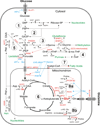
Figure 1
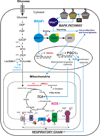
Figure 1. Metabolic rewiring in melanoma
Pathway end products are shown in green, factors affecting expression of metabolic genes or their regulators in blue (letters and arrows). OIS – oncogene induced senescence. Allosteric activation of PKM2 by serine is shown as red arrow. Metabolic enzyme and metabolite carrier names are in red. Glutamate as the major metabolite participating in both influx and outflow of glutamine carbon from the TCA cycle is shown in red bold letters and is also underlined. Dotted arrows indicate multistep reactions. Metabolic pathways: 1. Glycolysis, 2. Pentose phosphate pathway, 3. Serine and glycine biosynthesis, 4. Folate pathway, 5. Gluconeogenesis, 6. Tricarboxylic (TCA) acid cycle, 7. Fatty acid biosynthesis, 8. Proline biosynthesis: a – from glutamate, b – from arginine. Metabolites: G6P – glucose 6 phosphate, F6P – fructose 6 phosphate, GA3P – glyceraldehyde 3 phosphate, PEP – phosphoenolpyruvate, OAA – oxaloacetate, DHF – dihydrofolate, THF – tetrahydrofolate, me-THF - 5,10-methylene-THF, F-THF - 10-formyltetrahydrofolate, P5C – pyrroline-5 carboxylate, Genes: GLUT1 (SLC2A1) - facilitated glucose transporter member 1, PHGDH - phosphoglycerate dehydrogenase, SHMT1&2 – serine hydroxylmethyl transferases 1 (cytosolic) and 2 (mitochondrial), GCS – glycine cleavage system, DHFR – dihydrofolate reductase, PKM2 – pyruvate kinase isoform M2, PCK1 - phosphoenolpyruvate carboxykinase, LDHA – lactate dehydrogenase A, GOT1 and 2 – glutamate to oxaloacetate aminotransferase isoforms 1 and 2, MCP 1 and 2 – mitochondrial pyruvate carriers 1 and 2, PDH – pyruvate dehydrogenase complex, PDK1 – pyruvate dehydrogenase kinase, PDP2 - pyruvate dehydrogenase phosphatase 2, ASNS – asparagine synthetase, IDH1&2 – isocitate dehydrogenases 1 (cytosolic and peroxisomal) and 2 (mitochondrial), GLS – kidney type glutaminase, GLUD1 – glutamate dehydrogenase 1, GPT2 – glutamate to pyruvate aminotransferase 2, PYCR – pyrroline-5 carboxylate reductase: 1 – isoform 1, 2 – isoform 2, 3 – isoform 3, OAT – ornithine aminotransferase, SLC1A5 – glutamine plasma membrane carrier.
Figure 2
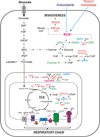
Figure 2. MAPK pathway activity, oncogenic BRAF and oxidative metabolism in melanoma
Oncogenic BRAF activity induces formation of ketone body acetoacetate, which enhances MAPK pathway activity by promoting binding between BRAFV600E and MEK1. Inhibition of oncogenic BRAF de-represses MITF expression leading to increased mitochondrial biogenesis and oxidative phosphorylation due to enhanced expression of the master mitochondrial regulator PGC1α, which also suppresses glycolysis. High PGC1α levels are critical for survival and tumorigenesis of a subset of melanomas relying heavily on oxidative metabolism by upregulating expression of ROS detoxification genes, thus mitigating oxidative stress. NRAS activation by mutations or loss of activity of the NF1 tumor suppressor can contribute to the activation of the MAPK pathway. Abbreviations: HMGCL - 3-hydroxy-3-methylglutaryl-CoA lyase, OXPHOS – genes encoding proteins participating in TCA cycle activity, oxidative phosphorylation and ATP production in mitochondria, ROS – reactive oxygen species, BRAFi – inhibitors of BRAFV600E activity, I – V – mitochondrial complexes 1 through 5.
Figure 3
Figure 3. ROS levels affect metastasis in melanoma
Distant metastasis causes oxidative stress in melanoma cells. Treatment with antioxidants promotes metastasis by enhancing GTP loading of RhoA GTPase and activating Rho-associated protein kinase (ROCK) thus inducing invasiveness. Activity of the folate pathway is important for mitigating oxidative stress by providing NADPH necessary for generation of reduced glutathione – a primary ROS detoxification agent. Abbreviations: GSH – reduced glutathione, GSSG – oxidized glutathione, MTHFD - methylenetetrahydrofolate reductase 1 (cytosolic) and 2 (mitochomdrial), SOD – superoxide dismutase, GPX – glutathione peroxidase, GSR – glutathione reductase, 10-formyltetrahydrofolate dehydrogenase

References

    1. The effect of vitamin E and beta carotene on the incidence of lung cancer and other cancers in male smokers. The Alpha-Tocopherol, Beta Carotene Cancer Prevention Study Group. N Engl J Med. 1994;330:1029–1035. - PubMed
    1. Abildgaard C, Guldberg P. Molecular drivers of cellular metabolic reprogramming in melanoma. Trends Mol Med. 2015;21:164–171. - PubMed
    1. Amelio I, Cutruzzola F, Antonov A, Agostini M, Melino G. Serine and glycine metabolism in cancer. Trends Biochem Sci. 2014;39:191–198. - PMC - PubMed
    1. Baenke F, Chaneton B, Smith M, Van Den Broek N, Hogan K, Tang H, et al. Resistance to BRAF inhibitors induces glutamine dependency in melanoma cells. Mol Oncol. 2015 - PMC - PubMed
    1. Bamford S, Dawson E, Forbes S, Clements J, Pettett R, Dogan A, et al. The COSMIC (Catalogue of Somatic Mutations in Cancer) database and website. Br J Cancer. 2004;91:355–358. - PMC - PubMed

MeSH terms