

Review
doi: 10.1084/jem.20151885.
Epub 2016 Jun 6.
Live cell imaging to understand monocyte, macrophage, and dendritic cell function in atherosclerosis
Affiliations
- PMID: 27270892
- PMCID: PMC4925021
- DOI: 10.1084/jem.20151885
Item in Clipboard
Review
Live cell imaging to understand monocyte, macrophage, and dendritic cell function in atherosclerosis
J Exp Med.
.
Abstract
Intravital imaging is an invaluable tool for understanding the function of cells in healthy and diseased tissues. It provides a window into dynamic processes that cannot be studied by other techniques. This review will cover the benefits and limitations of various techniques for labeling and imaging myeloid cells, with a special focus on imaging cells in atherosclerotic arteries. Although intravital imaging is a powerful tool for understanding cell function, it alone does not provide a complete picture of the cell. Other techniques, such as flow cytometry and transcriptomics, must be combined with intravital imaging to fully understand a cell's phenotype, lineage, and function.
© 2016 McArdle et al.
Figures
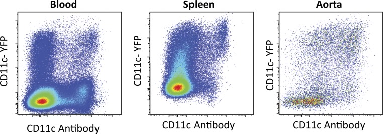
Representative data showing poor correlation between CD11c surface expression and YFP expression in Apoe−/− CD11cYFP mice in leukocytes the blood, spleen, and aorta via flow cytometry. All events gated for live CD45+ cells.
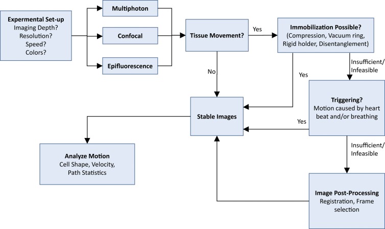
Decision flow chart for acquiring intravital videos. The choice of microscope depends mainly on the imaging depth (multiphoton better), resolution and colors (confocal better) or speed (epifluorescence better, see text for details). If tissue motion causes artifacts, mechanical immobilization, cardiac, and/or respiratory triggering, and image postprocessing can be used to stabilize the images, and then cell motion can be analyzed.
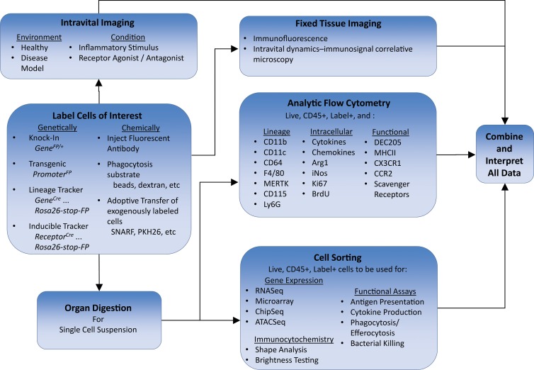
Determining phenotype and function of myeloid cells by combining intravital imaging with other techniques. Cells of interest are first labeled, either genetically with reporter mice or chemically, to be imaged in vivo. The labeled cells must also be analyzed with other techniques such as fixed tissue imaging, analytic flow cytometry for lineage and functional markers, functional assays, or gene expression. Finally, all data are combined for analysis and interpretation.
Comment in
-
Connecting Transcriptional and Functional Macrophage Heterogeneity in Atherosclerosis.Circ Res. 2019 Dec 6;125(12):1052-1054. doi: 10.1161/CIRCRESAHA.119.316168. Epub 2019 Dec 5. Circ Res. 2019. PMID: 31804906 Free PMC article. No abstract available.
References
Publication types
MeSH terms
Grants and funding
LinkOut - more resources
Full Text Sources
Other Literature Sources
Medical
