. 2016 Dec;17(9):1455-1466.
doi: 10.1111/mpp.12440. Epub 2016 Aug 21.

Dispensable chromosomes in Fusarium oxysporum f. sp. lycopersici

Affiliations

Dispensable chromosomes in Fusarium oxysporum f. sp. lycopersici

Ido Vlaardingerbroek et al. Mol Plant Pathol. 2016 Dec.

Abstract

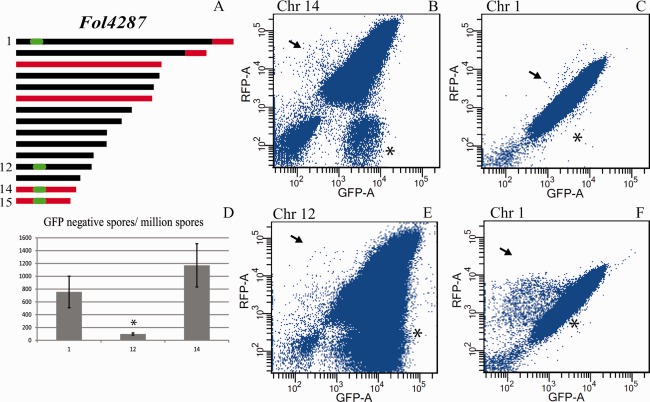
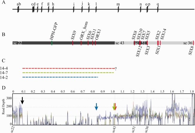
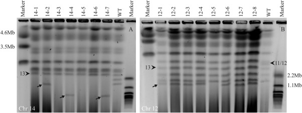
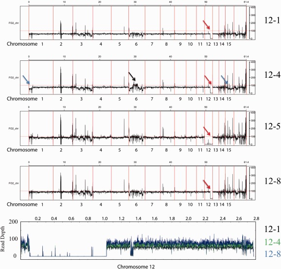
The genomes of many filamentous fungi consist of a 'core' part containing conserved genes essential for normal development as well as conditionally dispensable (CD) or lineage-specific (LS) chromosomes. In the plant-pathogenic fungus Fusarium oxysporum f. sp. lycopersici, one LS chromosome harbours effector genes that contribute to pathogenicity. We employed flow cytometry to select for events of spontaneous (partial) loss of either the two smallest LS chromosomes or two different core chromosomes. We determined the rate of spontaneous loss of the 'effector' LS chromosome in vitro at around 1 in 35 000 spores. In addition, a viable strain was obtained lacking chromosome 12, which is considered to be a part of the core genome. We also isolated strains carrying approximately 1-Mb deletions in the LS chromosomes and in the dispensable core chromosome. The large core chromosome 1 was never observed to sustain deletions over 200 kb. Whole-genome sequencing revealed that some of the sites at which the deletions occurred were the same in several independent strains obtained for the two chromosomes tested, indicating the existence of deletion hotspots. For the core chromosome, this deletion hotspot was the site of insertion of the marker used to select for loss events. Loss of the core chromosome did not affect pathogenicity, whereas loss of the effector chromosome led to a complete loss of pathogenicity.

Keywords: chromosome deletions; conditionally dispensable chromosomes; flow cytometry; pathogenic fungi.


Figures

Figure 1
Figure 1
Selection for loss of expression of a marker gene from selected chromosomes. (A) Schematic representation of core (black) and lineage‐specific (LS, red) parts of the genome of Fol4287. Locations targeted for GFP (green fluorescent protein gene) insertion are indicated in green. (B, C, E, F) Dot plots showing flow cytometry analysis of strains containing both GFP and RFP (red fluorescent protein gene). Axis labels indicate the detection channel used. Axis scale values are given in arbitrary units that were defined to fit the range of fluorescence in the biological samples. Each panel displays 1 × 10⁶ spores of a single culture with the GFP insertion in core chromosome 1 or 12 or LS chromosome 14 of Fol4287. Arrows indicate populations of spores that have lost GFP but not RFP, indicative of the loss of the associated genomic region. Asterisks indicate the populations that have lost RFP but not GFP. (D) Bar graph showing the number of GFP‐negative, RFP‐positive spores for strains expressing GFP from chromosome 1, 12 or 14, averaged over two repeats. This number is significantly (P = 1.6 × 10⁻⁵) lower for strains with GFP located on chromosome 12.
Figure 2
Figure 2
Location of markers and genes encoding proteins secreted in xylem (SIX) on positioned scaffolds of chromosome 14. (A) Location of primer pairs used to determine the size and location of the deletions in chromosome 14. The primer sequences are shown in Table S1 (see Supporting Information). (B) Location of genes for small secreted proteins (red bars) and of the insertion of the GFP (green fluorescent protein gene) marker (green bar) in chromosome 14. Alternating coloured bars indicate different supercontigs. Only positioned supercontigs are displayed. (C) Broken bars indicate the three deletions found, labelled with the strain number and colour coded. The deletion in 14‐4 continues until the end of supercontig 22, but does not continue in supercontig 43; the exact location of the deletion is unknown. All three strains lack the same five genes for in xylem‐secreted proteins. (D) Average read depth of genome sequences mapped to the reference for chromosome 14 for strains 14‐2 (blue line), 14‐7 (green line) and 14‐4 (red line). The black arrow marks the start of the gap in all three strains. The end of each gap is indicated by an arrow in the colour corresponding to the colour of the lines for each sample. Read depth for 14‐2 is higher for part of sc22, all of sc51 and part of sc36.
Figure 3
Figure 3
Karyotype analysis confirms partial or complete loss of chromosomes. (A) Contour‐clamped homogeneous electric field (CHEF) gel showing the size and number of chromosomes for strains which have lost chromosome 14 (14‐1, 14‐3, 14‐5 and 14‐6) or a region of chromosome 14 (14‐2, 14‐4 and 14‐7). Chromosome 13 is indicated with an arrowhead for reference. All strains lack the smallest chromosome from the wild‐type (WT) strain (chromosome 15) as well as the normal sized chromosome 14. Strains 14‐2, 14‐4 and 14‐7 have a smaller chromosome absent in the WT strain, indicated with arrows. (B) CHEF gel analysis of strains with partial or complete loss of chromosome 12 (labelled 12‐1–12‐8). Chromosomes 11 and 12, which run closely together, are indicated with an arrowhead. In all the selected strains without GFP (green fluorescent protein gene), the band corresponding to chromosome 12 is missing. Below chromosome 11 in these strains are chromosomes 13–15, in most cases followed by a novel chromosome. This presumed remainder of chromosome 12 is indicated with an arrow in 12‐1. This chromosome is absent in 12‐5, suggesting the complete loss of chromosome 12 in this strain.
Figure 4
Figure 4
Average read depth reveals duplication and loss events. Average read depth graphs for strains that lost chromosome 12. Strains 12‐1, 12‐4 and 12‐8 all appear to have the same deletion in chromosome 12, indicated by red arrows. The average read depth mapped against the supercontig representing most of chromosome 12 from our own assembly is shown in the bottom panel (12‐1 in black, 12‐4 in green and 12‐8 in blue). Here, we can clearly see that all three strains lack the same region. Strain 12‐4 has a duplicated region on chromosome 6 relative to the reference genome, indicated by a black arrow. This duplication event appears to be very similar to that shown for strain 14‐2 in Fig. 5. Strain 12‐4 has a lower read depth for the lineage‐specific (LS) region of chromosomes 1 and 15 compared with strains 12‐1 and 12‐8, indicated by blue arrows.
Figure 5
Figure 5
Average read depth reveals duplication and loss events. The panels show the average read depth for the strain from which chromosome 14 was lost completely (14‐1) and three strains which have a 0.9‐Mb deletion in chromosome 14, indicated by green arrows. Strain 14‐2 has a smaller deletion than 14‐4 and 14‐7. Strain 14‐2 has a duplicated region on chromosome 6 relative to the reference genome, indicated by a black arrow.
Figure 6
Figure 6
Chromosome 12 and a large region of chromosome 14 are dispensable for pathogenicity. (A) Plants infected with strain 14‐6, which lacks chromosome 14, are completely healthy. (B) Plants infected with strain 14‐7, from which part of chromosome 14 is lost, show disease symptoms similar to those of the wild‐type and parental strains. (C) Distribution of disease index (DI) for strains which have lost part or all of chromosome 14. DI was scored in the range 0–4 based on the number of brown vessels and macroscopic effects, as described previously (Rep et al., 2004). Twenty plants were used per strain. The experiment was performed twice with similar results. 1‐1 indicates 14HG1+RFP1 and 1‐2 indicates 14HG1+RFP2; these are the strains from which the other strains were selected and they show disease severity similar to that of the wild‐type Fol4287 (despite the absence of chromosome 15). 14‐7 was selected from a culture of 14HG1+RFP2, whereas the other strains all originate from 14HG1+RFP1 cultures. The pathogenicity of strains 14‐2, 14‐4 and 14‐7, which have all sustained a deletion in chromosome 14, is not significantly different from that of the wild‐type. Strains completely lacking chromosome 14 have lost pathogenicity. (D) DI for five independent strains (labelled C1–C5) derived from chromosome 14‐lacking strains complemented with chromosome 14 through horizontal chromosome transfer. All strains show a regain of pathogenicity. (E, F) Photographs of plants infected with strains 12‐1 and 12‐4 which have lost part of chromosome 12. (G) DI for strains lacking part or all of chromosome 12. +RFP1 is strain 12HG1+RFP1 from which strains 12‐1–12‐5 originate; strains 12‐6–12‐8 originate from 12HG1+RFP2. (H) Average plant weight for strains that have lost chromosome 14 or sustained large deletions. No significant differences were observed.

References

    1. Balesdent, M. , Fudal, I. , Ollivier, B. , Bally, P. , Grandaubert, J. , Eber, F. , Chèvre, A. , Leflon, M. and Rouxel, T. (2013) The dispensable chromosome of Leptosphaeria maculans shelters an effector gene conferring avirulence towards Brassica rapa . New Phytol. 198, 887–898. - PubMed
    1. Chuma, I. , Isobe, C. , Hotta, Y. , Ibaragi, K. , Futamata, N. , Kusaba, M. , Yoshida, K. , Terauchi, R. , Fujita, Y. and Nakayashiki, H. (2011) Multiple translocation of the AVR‐pita effector gene among chromosomes of the rice blast fungus Magnaporthe oryzae and related species. PLoS Pathog. 7, e1002147. - PMC - PubMed
    1. Coleman, J.J. , Rounsley, S.D. , Rodriguez‐Carres, M. , Kuo, A. , Wasmann, C.C. , Grimwood, J. , Schmutz, J. , Taga, M. , White, G.J. and Zhou, S. (2009) The genome of Nectria haematococca: contribution of supernumerary chromosomes to gene expansion. PLoS Genet. 5, e1000618. - PMC - PubMed
    1. Covert, S.F. (1998) Supernumerary chromosomes in filamentous fungi. Curr. Genet. 33, 311–319. - PubMed
    1. Croll, D. and McDonald, B.A. (2012) The accessory genome as a cradle for adaptive evolution in pathogens. PLoS Pathog. 8, e1002608. - PMC - PubMed

Publication types

LinkOut - more resources