Review
. 2016 Jun 2;8(6):156.
doi: 10.3390/v8060156.

The Role of microRNAs in the Pathogenesis of Herpesvirus Infection

Affiliations
Review

The Role of microRNAs in the Pathogenesis of Herpesvirus Infection

Diogo Piedade et al. Viruses. .

Abstract

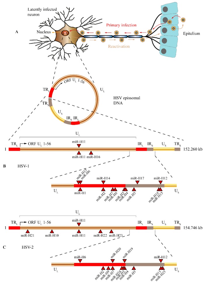
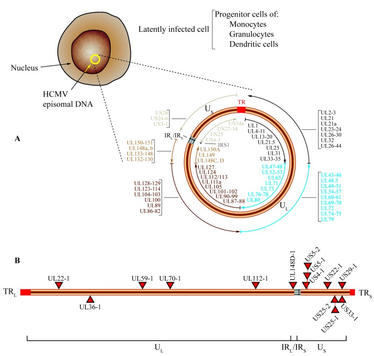
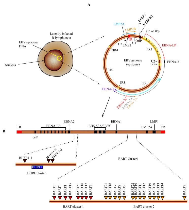
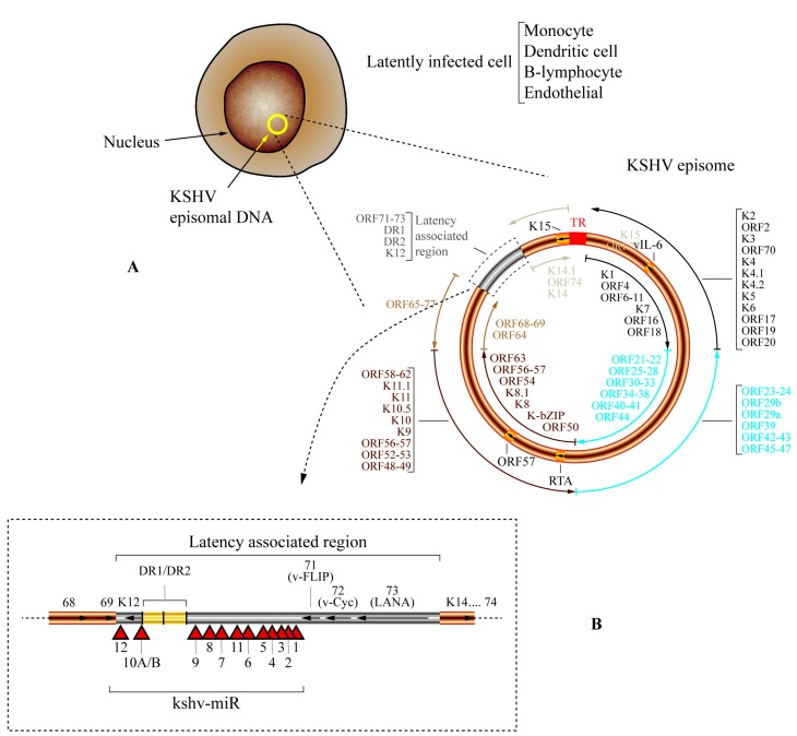
MicroRNAs (miRNAs) are small non-coding RNAs important in gene regulation. They are able to regulate mRNA translation through base-pair complementarity. Cellular miRNAs have been involved in the regulation of nearly all cellular pathways, and their deregulation has been associated with several diseases such as cancer. Given the importance of microRNAs to cell homeostasis, it is no surprise that viruses have evolved to take advantage of this cellular pathway. Viruses have been reported to be able to encode and express functional viral microRNAs that target both viral and cellular transcripts. Moreover, viral inhibition of key proteins from the microRNA pathway and important changes in cellular microRNA pool have been reported upon viral infection. In addition, viruses have developed multiple mechanisms to avoid being targeted by cellular microRNAs. This complex interaction between host and viruses to control the microRNA pathway usually favors viral infection and persistence by either reducing immune detection, avoiding apoptosis, promoting cell growth, or promoting lytic or latent infection. One of the best examples of this virus-host-microRNA interplay emanates from members of the Herperviridae family, namely the herpes simplex virus type 1 and type 2 (HSV-1 and HSV-2), human cytomegalovirus (HCMV), human herpesvirus 8 (HHV-8), and the Epstein-Barr virus (EBV). In this review, we will focus on the general functions of microRNAs and the interactions between herpesviruses, human hosts, and microRNAs and will delve into the related mechanisms that contribute to infection and pathogenesis.

Keywords: herpesvirus; immune evasion; latency; microRNAs; oncogenesis; pathogenesis.


Figures

Figure 1
Figure 1
Viral genomic DNA structure and location of microRNAs (miRNAs) encoded by herpes simplex virus type 1 and type 2 (HSV-1 and HSV-2). (A) Episomal DNA in a latently infected neuron. After primary infection, HSV-1 or HSV-2 virions migrate from oral or genital mucosa to a sensitive neuron that enervates those tissues establishing a latent infection. The migration through neuronal dendrites occurs by a retrograde microtubule-associated transport, allowing HSV to reach nerve cell body. As a response to several exogenous or endogenous stimuli, the latent virus is reactivated and viral particles are anterograde transported to the mucosa where they infect epithelial cells allowing viral transmission. During latency, the HSV genome is maintained as a circular episome (represented as a red circle inside the nucleus). The structure and prototypic arrangement of the HSV episome is presented. UL (copper) and US (gold) represent the long and short components of the viral genome. TRL and IRL (in red) represent the repeat sequences flanking UL and TRS, and IRS (in light brown) represent the repeat sequences flanking US. Genomic location of miRNAs encoded by HSV-1 (B) and HSV-2 (C). A linear representation of both HSV-1 and HSV-2 genomes are shown, and the locations of miRNA precursors encoded by each virus are denoted by red triangles. The regions of the internal repeat sequences (IRL and IRS) are expanded in the bottom of each genome. miRNAs shown below the line representing viral double-strand DNA are transcribed in an antisense direction (from left to right), while those shown above the line are transcribed in the opposite orientation.
Figure 2
Figure 2
Genome organization and location of miRNA encoded by human cytomegalovirus (HCMV). (A) After primary infection HCMV establishes a latent infection in progenitor cells of monocytes, dendritic cells, and granulocytes. During latency, the HCMV genome is maintained as a circular episome (represented as a yellow circle inside the nucleus). The general structure of episomal DNA is presented, and the relative positions of the open reading frames (ORFs) are shown. UL and US represent the long and short components of the viral genome, TR (red) denotes the terminal repeats, and IRL/IRS (grey) represents the internal repeats of the long and short components of the HCMV genome. The outer circular arrows refer to the ORFs that are transcribed in antisense orientation, while the inner circular arrows refer to those that are transcribed in the opposite orientation. To facilitate the representation of the different ORFs and their relative location, the outer and inner circular arrows are presented in different colors; the black circular arrows represent those ORFs encoded between 12 and 58 kb of the HCMV genome, the blue circular arrows represent ORFs encoded between 59 and 118 kb, the dark brown circular arrows denote the ORFs encoded between 119 and 177 kb, the light brown circular arrows refer to the ORFs located between 178 kb and the IRL/IRS region, and finally the light grey circular arrows represent the ORFs that are encoded after the IRL/IRS region; (B) Genomic location of miRNAs encoded by HCMV. A linear representation of the HCMV genome is shown and the locations of miRNA precursors are denoted by red triangles. miRNAs shown below the line representing viral double-strand DNA are transcribed in antisense direction (from left to right), while those shown above are transcribed in the opposite orientation.
Figure 3
Figure 3
Genome organization and location of miRNAs encoded by the Epstein–Barr virus (EBV). (A) After primary infection, EBV establishes a latent infection in B-lymphocytes. During latency, the EBV genome is maintained as a circular episome (represented as a yellow circle inside the nucleus). The general structure of episomal DNA is presented, and the relative positions of the latency-associated genes on the viral episome are shown. The origin of plasmid replication (oriP) is shown in brown. The outer circle represents the coding regions of the latent proteins transcribed in antisense (LMP1, black arrow) and sense orientation (EBNA-LP, red; EBNA2, black; EBNA3A, light brown; EBNA3B, light grey; EBNA3C, red; EBNA1, purple; LMP2A, blue; LMP2B, orange). EBNA-LP is transcribed from variable numbers of repetitive exons (denoted by black lines inside the IR1 region). The highly transcribed non-polyadenylated RNAs EBER1 and EBER2 are represented in the top of the diagram. The outer long circular arrow represents the EBV transcript during the latency III program where all the EBNA genes are transcribed from Cp or Wp promoters. The inner blue arrow line represents the EBNA1 transcript originated from the Qp promoter during latency I and latency II programs. U1-U5 refers to largely unique DNA domains while IR1-IR4 represents internal repetitive DNA domains. TR (represented in red) denotes the terminal repeat of EBV DNA; (B) Genomic location of miRNAs encoded by EBV. A linear representation of EBV genome is shown and the positions of the main latent genes are represented by black boxes; a dark brown box denotes oriP position. The regions of the BHRF cluster and BART clusters 1 and 2 are expanded in the bottom of the diagram. The locations of miRNA precursors are denoted by brown (BHRF cluster), red (BART cluster 1) and yellow (BART cluster 2) triangles.
Figure 4
Figure 4
Genome organization and location of miRNA encoded by Kaposi’s sarcoma-associated herpesvirus (KSHV) (HHV-8). (A) After primary infection, KSHV establishes a latent infection in monocytes, dendritic cells, B-lymphocytes, and endothelial cells. During latency, the KSHV genome is maintained as a circular episome (represented as a yellow circle inside the nucleus). The general structure of episomal DNA is presented, and the relative positions of the open reading frames (ORFs) are shown. The grey box represents the latency-associated region. The terminal repeat (TR) region is represented by a red box, and the ORFs encoding for viral interleukin 6 (vIL-6), the replication and transcription activator (RTA), ORF57, and K15 are represented by light orange boxes. The outer circular arrows refer to the ORFs that are transcribed in antisense orientation, while the inner circular arrows refer to those that are transcribed in the opposite orientation. To facilitate the representation of the different ORFs and their relative location, and the outer and inner circular arrows are presented in different colors (black, light blue, brown, light brown and light grey); (B) Genomic location of miRNAs encoded by KSHV (kshv-miR). A linear representation of the latency-associated region (grey box) of the KSHV genome is shown, and the locations of miRNA precursors are denoted by red triangles. The positions of ORFs 68, 69, K12, 71 (v-FLIP), 72 (v-Cyc), 73 (LANA), K14, and 74 are also shown; arrows inside each ORF indicate the direction of transcription. The two adjacent yellow boxes represent the DR1/DR2 region.

References

    1. Bushati N., Cohen S.M. microRNA functions. Annu. Rev. Cell. Dev. Biol. 2007;23:175–205. doi: 10.1146/annurev.cellbio.23.090506.123406. - DOI - PubMed
    1. Kloosterman W.P., Plasterk R.H.A. The Diverse Functions of MicroRNAs in Animal Development and Disease. Dev. Cell. 2006;11:441–450. doi: 10.1016/j.devcel.2006.09.009. - DOI - PubMed
    1. Doench J.G., Petersen C.P., Sharp P.A. siRNAs can function as miRNAs. Genes Dev. 2003;17:438–442. doi: 10.1101/gad.1064703. - DOI - PMC - PubMed
    1. Bartel D.P. MicroRNAs: Genomics, Biogenesis, Mechanism, and Function. Cell. 2004;116:281–297. doi: 10.1016/S0092-8674(04)00045-5. - DOI - PubMed
    1. Lee R.C., Feinbaum R.L., Ambros V. The C. elegans heterochronic gene lin-4 encodes small RNAs with antisense complementarity to lin-14. Cell. 1993;75:843–854. doi: 10.1016/0092-8674(93)90529-Y. - DOI - PubMed

MeSH terms

LinkOut - more resources