Review
. 2016 Aug;27(7-8):367-80.
doi: 10.1007/s00335-016-9650-y. Epub 2016 Jun 6.

Genetics of aging bone

Affiliations
Review

Genetics of aging bone

Douglas J Adams et al. Mamm Genome. 2016 Aug.

Abstract

With aging, the skeleton experiences a number of changes, which include reductions in mass and changes in matrix composition, leading to fragility and ultimately an increase of fracture risk. A number of aspects of bone physiology are controlled by genetic factors, including peak bone mass, bone shape, and composition; however, forward genetic studies in humans have largely concentrated on clinically available measures such as bone mineral density (BMD). Forward genetic studies in rodents have also heavily focused on BMD; however, investigations of direct measures of bone strength, size, and shape have also been conducted. Overwhelmingly, these studies of the genetics of bone strength have identified loci that modulate strength via influencing bone size, and may not impact the matrix material properties of bone. Many of the rodent forward genetic studies lacked sufficient mapping resolution for candidate gene identification; however, newer studies using genetic mapping populations such as Advanced Intercrosses and the Collaborative Cross appear to have overcome this issue and show promise for future studies. The majority of the genetic mapping studies conducted to date have focused on younger animals and thus an understanding of the genetic control of age-related bone loss represents a key gap in knowledge.


Figures
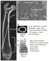
Figure 1
Figure 1. Organization of long bones
A. The structure of the mouse femur is shown by longitudinal section of a three-dimensional X-ray microtomography image, showing prominent cortical and trabecular bone regions. B. Cortical morphometry, as measured by microtomography, typically is defined at mid-diaphysis by measurement of total area, marrow area, cortical area and thickness, major and minor dimensions, and geometric indices of flexural and torsional stiffness and strength. Strength of long bones, as measured by flexure testing, usually involves breaking the bone at the mid-shaft. C. Trabecular morphometry, also measured by microtomography, is defined by measurement of trabecular bone volume as a percent of total volume inside the cortical envelope, trabecular thickness and number. Other measures describing geometry typically are collected, as are listed in the image. D. A hematoxylin and eosin stained section from the distal femur (top) shows cortical bone to the left with a trabeculum in the middle. Osteoclasts and osteoblasts are found on bone surfaces whereas osteocytes are embedded in the bone. The lamellar structure of bone can be seen as bands of darker versus weaker staining.
Figure 2
Figure 2. The influence of bone size and composition on density and strength
Idealized diaphyseal cross-sections of equivalent area-projected bone mineral density. Although BMD is numerically identical, the amount of cortical bone, thickness of cortex, and geometrical strength in bending or twisting are not equivalent. Further BMD is not the same as Tissue Density (see Figure 5). Note the resemblance to actual mouse femur mid-diaphysis cross-sections shown in Figure 4.
Figure 3
Figure 3. Trabecular bone volume fraction (BV/TV) in DO mice
BV/TV was measured in the distal femur in 6, 12 and 18 month old male and female DO mice by μCT by using standard methods. Even at six months of age some of the DO mice presented with a BV/TV of zero, indicating that all trabecular bone had been resorbed in these animals. The BV/TV was lowest in the oldest cohort of animals, which is reminiscent of the decrease in trabecular bone seen with aging in humans.
Figure 4
Figure 4. Representative images of DO mice femurs as captured by microcomputed tomography
Cross-sections of the mid-diaphysis (A) and the distal trabecular compartment (B) of femurs. All images are from 6 month old females. Images from A and B are not from the same animals. These representative animals highlight the vast diversity in bone phenotypes observed in this genetic mapping population.
Figure 5
Figure 5. Bone size effect on bone mineral density (BMD) measures made by dual X-ray absorptiometry (DXA)
Whole body BMD (A) and bone area (B) were measured by DXA in 17 week old male WSB/EiJ and NZO/H1LtJ mice. The head region was excluded from these measures. The density of the cortical bone of the tibia was measured by microcomputed tomography in these same mice (C). As expected, BMD was higher in the NZO/H1LtJ mice, which is largely a function of the much larger skeletal size of these mice. However, the true matrix mineral density of the cortical bone of these mice is not different. These data highlight that what is considered BMD for a whole animal does not reflect the true density of the actual bone tissue. The difference between BMD and matrix mineral density is an important consideration when interpreting candidate genes for BMD.

References

    1. Leali PT, Muresu F, Melis A, Ruggiu A, Zachos A, Doria C. Skeletal fragility definition. Clin Cases Miner Bone Metab. 2011 May;8(2):11–3. Epub 2012/03/31. - PMC - PubMed
    1. Blume SW, Curtis JR. Medical costs of osteoporosis in the elderly Medicare population. Osteoporos Int. 2011 Jun;22(6):1835–44. - PMC - PubMed
    1. von Friesendorff M, McGuigan FE, Besjakov J, Akesson K. Hip fracture in men-survival and subsequent fractures: a cohort study with 22-year follow-up. J Am Geriatr Soc. 2011 May;59(5):806–13. - PubMed
    1. Willson T, Nelson SD, Newbold J, Nelson RE, LaFleur J. The clinical epidemiology of male osteoporosis: a review of the recent literature. Clin Epidemiol. 2015;7:65–76. - PMC - PubMed
    1. Brauer CA, Coca-Perraillon M, Cutler DM, Rosen AB. Incidence and mortality of hip fractures in the United States. JAMA. 2009 Oct 14;302(14):1573–9. - PMC - PubMed

Publication types