. 2016 Jun 8:6:27557.
doi: 10.1038/srep27557.

Disruption of Slc52a3 gene causes neonatal lethality with riboflavin deficiency in mice

Affiliations

Disruption of Slc52a3 gene causes neonatal lethality with riboflavin deficiency in mice

Hiroki Yoshimatsu et al. Sci Rep. .

Abstract

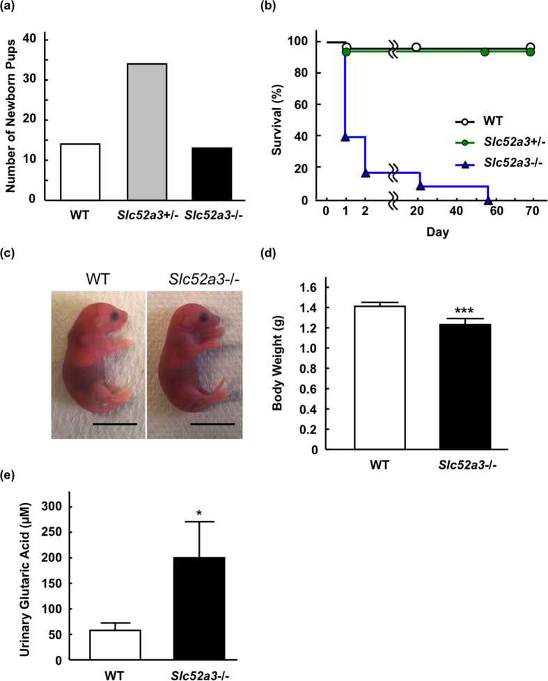
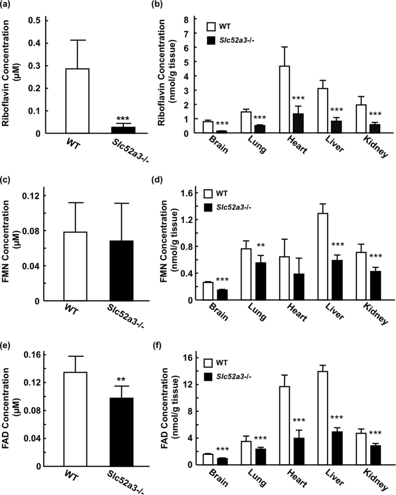
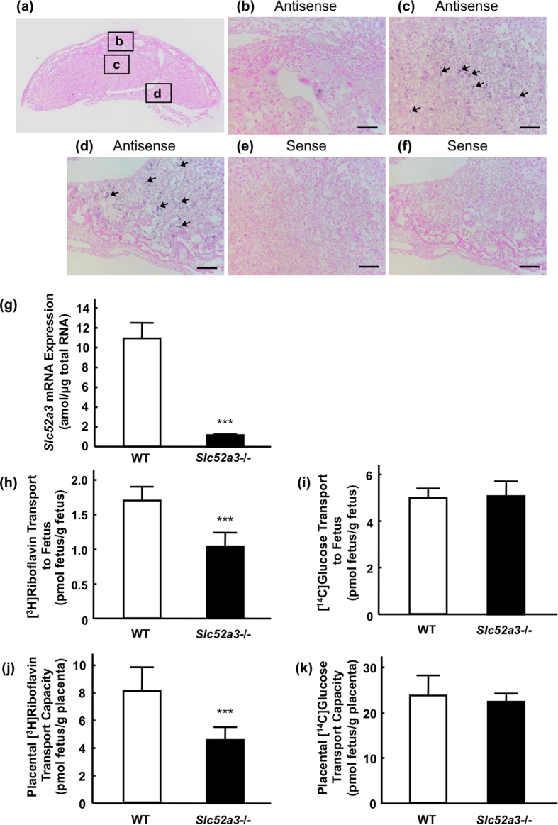
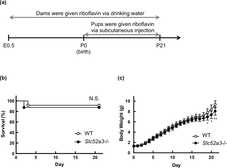
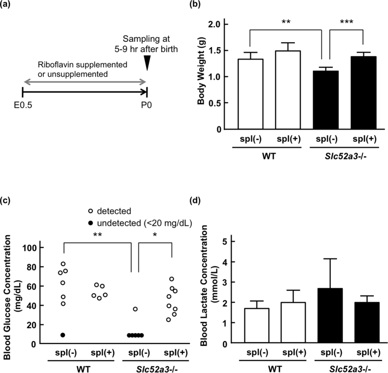
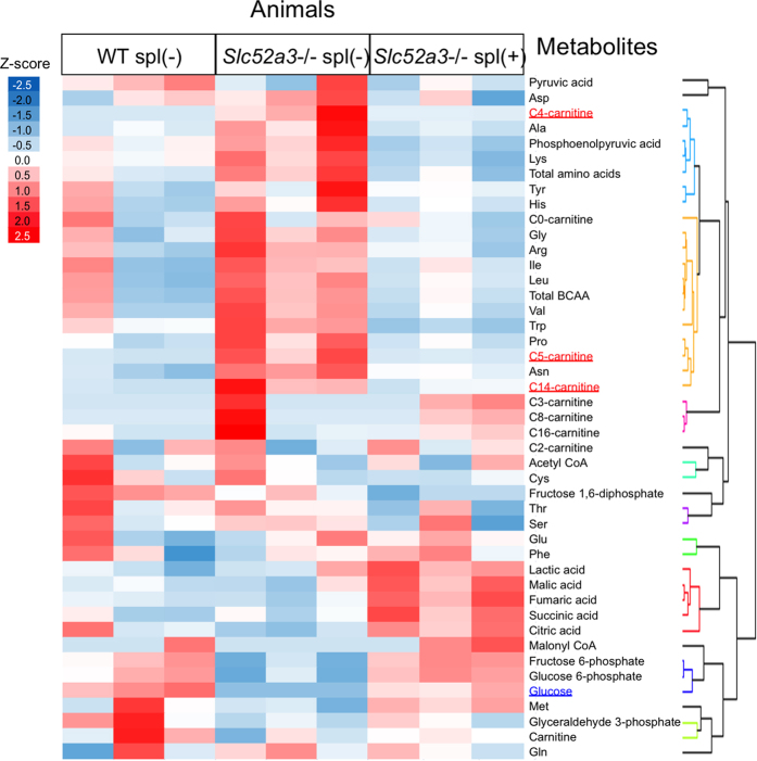
Homeostasis of riboflavin should be maintained by transporters. Previous in vitro studies have elucidated basic information about riboflavin transporter RFVT3 encoded by SLC52A3 gene. However, the contribution of RFVT3 to the maintenance of riboflavin homeostasis and the significance in vivo remain unclear. Here, we investigated the physiological role of RFVT3 using Slc52a3 knockout (Slc52a3-/-) mice. Most Slc52a3-/- mice died with hyperlipidemia and hypoglycemia within 48 hr after birth. The plasma and tissue riboflavin concentrations in Slc52a3-/- mice at postnatal day 0 were dramatically lower than those in wild-type (WT) littermates. Slc52a3-/- fetuses showed a lower capacity of placental riboflavin transport compared with WT fetuses. Riboflavin supplement during pregnancy and after birth reduced neonatal death and metabolic disorders. To our knowledge, this is the first report to indicate that Rfvt3 contributes to placental riboflavin transport, and that disruption of Slc52a3 gene caused neonatal mortality with hyperlipidemia and hypoglycemia owing to riboflavin deficiency.


Figures

Figure 1
Figure 1. Phenotypic analyses of Slc52a3−/− mice.
(a) Genotypic distribution immediately after birth (n = 63 from 8 litters). (b) Kaplan-Meier survival curves for WT (n = 14; open circles, black line), Slc52a3+/− (n = 34; closed circles, green line) and Slc52a3−/− (n = 13; closed triangles, blue line) littermates. (c) Gross appearance of whole body of WT, Slc52a3+/− and Slc52a3−/− newborn pups at postnatal day 0 (P0). (d) Body weight of WT, Slc52a3+/− and Slc52a3−/− littermates at P0. (e) Urinary glutaric acid levels in WT and Slc52a3−/− littermates at P0. Each scale bar represents 1 cm length. Each column or point represents the mean ± SD. Values where *P < 0.05, or ***P < 0.001, significantly different from WT.
Figure 2
Figure 2. Riboflavin, FMN, and FAD levels in plasma and tissues from WT and Slc52a3−/− mice.
Plasma and tissues were obtained from WT (n = 14) and Slc52a3−/− (n = 13) littermates at postnatal day 0 (P0) immediately after birth. Plasma concentrations of riboflavin (a), FMN (c) and FAD (e) were measured by HPLC. Tissue levels of riboflavin (b), FMN (d) and FAD (f) were also determined. Each column represents the mean ± SD. Values where **P < 0.01, ***P < 0.001, significantly different from WT.
Figure 3
Figure 3. Involvement of Rfvt3 in the placental riboflavin transport.
(a) mRNA expression of Slc52a3 was examined by in situ hybridization. The insets indicate the magnification region. (b–d) Higher magnification images corresponding the solid flames in (a): deciduum (b), junctional zone (c) and labyrinth zone (d). The blue signals were positive staining for Slc52a3 (arrows). Hybridization with a sense probe for Slc52a3 (negative control) in the junctional zone (e) and labyrinth zone (f). Scale bar: 100 μm. (g) Slc52a3 mRNA expression in the placentas from WT and Slc52a3−/− fetuses derived from Slc52a3+/− dams. Total RNA isolated from the placentas from WT and Slc52a3−/− fetuses was reverse-transcribed and mRNA level of Rfvt3 was determined by real-time PCR. Each column represents the mean ± SD (WT, n = 6; Slc52a3−/−, n = 6 from 2 litters). (h,i) Placental transport of [3H]riboflavin in WT and Slc52a3−/− fetuses. Slc52a3+/− mice were mated, and [3H]riboflavin was administered intravenously to pregnant dams at gestation day 16.5. Five minutes after administration, fetuses and placentas were collected. Placental transport of [3H]riboflavin was expressed per gram of placenta (h) or per gram of fetus (i). Placental transport of [14C]glucose was also examined (j,k). Each column represents the mean ± SD (WT, n = 7; Slc52a3−/−, n = 8 from 3 litters). Values where ***P < 0.001 were significantly different from WT.
Figure 4
Figure 4. Survival of Slc52a3−/− mice with riboflavin supplementation.
(a) Protocol for the riboflavin supplementation. Slc52a3+/− mice were mated, and pregnant dams were supplemented with 50 mg/L of riboflavin in drinking water ad libitum from gestational day 0 to 3 weeks postpartum. Newborn pups were also administered 0.75 mg/kg riboflavin subcutaneously once a day until weaning at the postnatal day 21. Kaplan-Meier survival (b) and body weights (c) for WT and Slc52a3−/− mice with riboflavin administration. The body weight values are exressed as the mean ± SD for mice that were alive at each time point (WT, n = 12; Slc52a3−/−, n = 8 from 4 litters).
Figure 5
Figure 5. Blood glucose and lactate levels in Slc52a3−/− neonatal mice with or without riboflavin supplementation.
(a) Protocol for the riboflavin supplementation. In the riboflavin-supplemented group (spl (+)), the pregnant dams were given 50 mg/L of riboflavin in drinking water ad libitum from gestational day 0. Postnatal day 0 (P0) pups were collected at 5–9 hr after birth. (b) Body weights of WT and Slc52a3−/− pups. (c) Blood glucose levels in WT and Slc52a3−/− pups. Closed circles represent undetected data (<20 mg/dL). (d) Blood lactate levels in WT and Slc52a3−/− pups. Each column represents the mean ± SD (WT spl(−), n = 7; WT spl(+), n = 5; Slc52a3−/− spl(−), n = 6; Slc52a3−/− spl(+), n = 8). Values where *P < 0.05, **P < 0.01, or ***P < 0.001 indicates significant difference.
Figure 6
Figure 6. Metabolome analysis in Slc52a3−/− neonatal mice.
Hierarchical clustering of targeted metabolomics data. Amino acids, metabolites of glycolysis and TCA cycle in liver, plasma acylcarnitine, and blood glucose in WT, Slc52a3−/− pups (Slc52a3−/− spl(−)) and riboflavin-supplemented Slc52a2−/− pups (Slc52a3−/− spl(+)) are listed on the abscissa. The color in the heat map reflects the relative metabolite abundance level according to the z-score.

References

    1. Depeint F., Bruce W. R., Shangari N., Mehta R. & O’Brien P. J. Mitochondrial function and toxicity: role of the B vitamin family on mitochondrial energy metabolism. Chem Biol Interact 163, 94–112 (2006). - PubMed
    1. Powers H. J. Riboflavin (vitamin B-2) and health. Am J Clin Nutr 77, 1352–1360 (2003). - PubMed
    1. Robitaille J., Carmichael S. L., Shaw G. M. & Olney R. S. Maternal nutrient intake and risks for transverse and longitudinal limb deficiencies: data from the National Birth Defects Prevention Study, 1997–2003. Birth Defects Res A Clin Mol Teratol 85, 773–779 (2009). - PubMed
    1. Smedts H. P. et al. Maternal intake of fat, riboflavin and nicotinamide and the risk of having offspring with congenital heart defects. Eur J Nutr 47, 357–365 (2008). - PubMed
    1. Foraker A. B., Khantwal C. M. & Swaan P. W. Current perspectives on the cellular uptake and trafficking of riboflavin. Adv Drug Deliv Rev 55, 1467–1483 (2003). - PubMed

Publication types

MeSH terms