. 2016 Aug;12(8):586-92.
doi: 10.1038/nchembio.2098. Epub 2016 Jun 6.

Copper regulates cyclic-AMP-dependent lipolysis

Affiliations

Copper regulates cyclic-AMP-dependent lipolysis

Lakshmi Krishnamoorthy et al. Nat Chem Biol. 2016 Aug.

Abstract

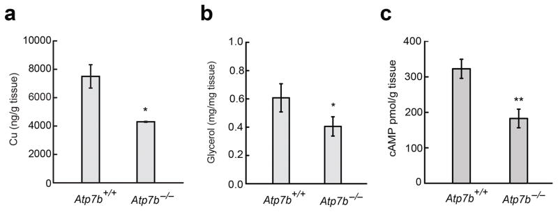
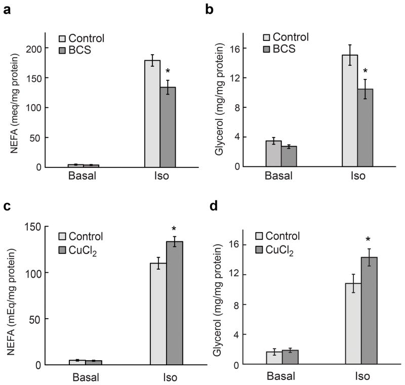
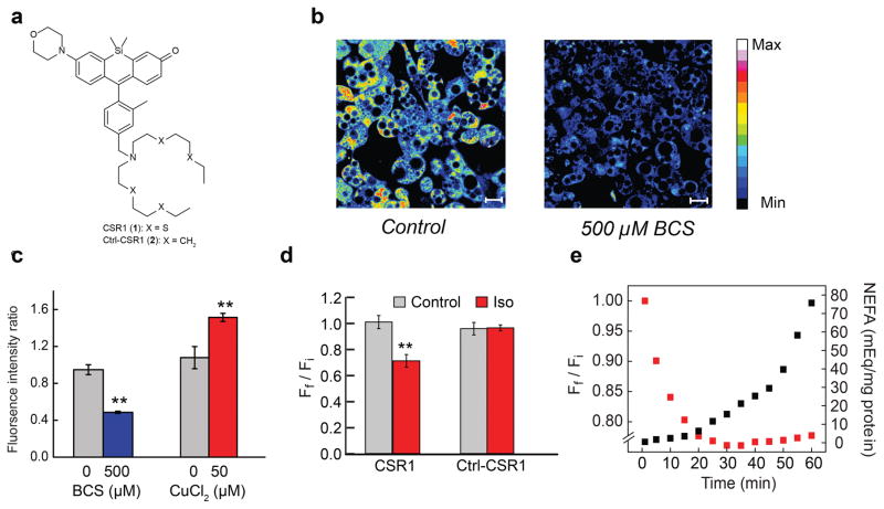
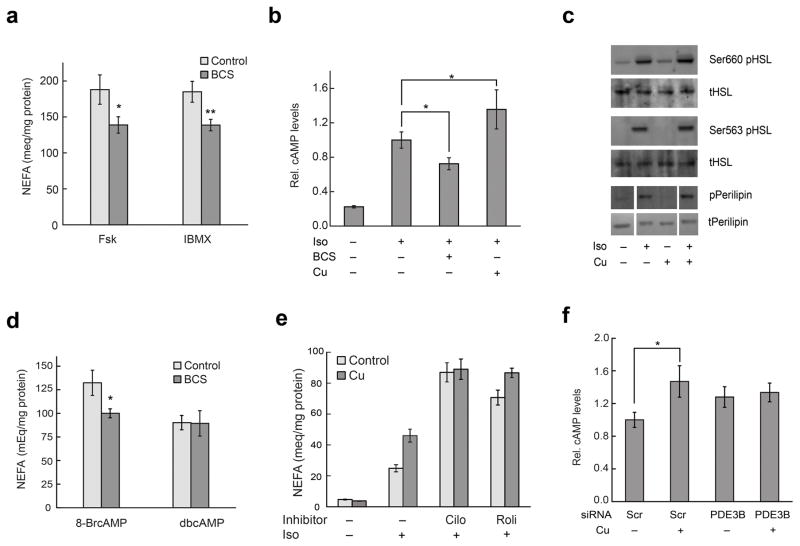
Cell signaling relies extensively on dynamic pools of redox-inactive metal ions such as sodium, potassium, calcium and zinc, but their redox-active transition metal counterparts such as copper and iron have been studied primarily as static enzyme cofactors. Here we report that copper is an endogenous regulator of lipolysis, the breakdown of fat, which is an essential process in maintaining body weight and energy stores. Using a mouse model of genetic copper misregulation, in combination with pharmacological alterations in copper status and imaging studies in a 3T3-L1 white adipocyte model, we found that copper regulates lipolysis at the level of the second messenger, cyclic AMP (cAMP), by altering the activity of the cAMP-degrading phosphodiesterase PDE3B. Biochemical studies of the copper-PDE3B interaction establish copper-dependent inhibition of enzyme activity and identify a key conserved cysteine residue in a PDE3-specific loop that is essential for the observed copper-dependent lipolytic phenotype.


Conflict of interest statement

Competing Financial Interests

The authors declare no competing financial interests.

Figures

Fig. 1
Fig. 1
Genetically induced copper misregulation affects lipid metabolism in vivo. (a) Copper content of WAT from Atp7b+/+ and Atp7b−/− mice. (n = 3). (b) Glycerol release from Iso-stimulated explant WAT from Atp7b+/− and Atp7b−/− mice (Het n = 5, KO n = 3) (c) cAMP content of Iso-stimulated explant WAT from Atp7b+/− and Atp7b−/− mice (Het n = 5, KO n = 4). Values are shown as mean ± s.e.m. * p < 0.05. ** p < 0.01.
Fig. 2
Fig. 2
Labile copper pools alter cAMP-dependent lipolysis. For copper chelation experiments (a,b), 3T3-L1 adipocytes were treated with 500 μM BCS for 20 h in DMEM with 2% BSA. For copper supplementation experiments (c,d), cells were incubated for 20 h in DMEM with 2% BSA followed by treatment with 50 μM copper for 1 h in DMEM alone; copper supplementation was performed in the absence of BSA as BSA binds Cu2+ tightly. For both treatments, cells were then stimulated with 100 nM Iso for 60 min in DMEM with 2% BSA. Culture media were harvested for analysis of (a,c) NEFA and (b,d) glycerol release. Values are shown as mean ± s.e.m. (n = 3). * p < 0.05.
Fig. 3
Fig. 3
Molecular imaging reveals that lipolysis alters labile copper. (a) Structure of CSR1. (b) Images of 3T3-L1 cells supplemented with vehicle or 500 μM BCS in the growth medium for 24 h, stained with 2 μM CSR1 for 10 min at 37 °C in DMEM and then imaged. Scale bar; 20 μm. (c) Quantification of images of 3T3-L1 cells stained with 2 μM CSR1 in DMEM at 37 °C under conditions of copper-supplementation and BCS treatment. The ratio of fluorescent intensity of control cells and treated cells is shown. Values are shown as mean ± s.e.m. (n = 3). (d) 3T3-L1 adipocytes were stained with CSR1 or Ctrl-CSR1 and then stimulated with 100 nM Iso for 60 min. Quantification of images obtained represent Ff /Fi values for control and Iso-treated cells stained with CSR1 or Ctrl-CSR1, respectively.(e) Graph displays data points for concomitant changes in CSR1 fluorescence (red squares) and NEFA release (black squares) obtained every 5 min during lipolysis over 60 min. Values are shown as mean ± s.e.m. (n = 3). ** p < 0.01.
Fig. 4
Fig. 4
Labile copper pools alter cAMP levels by regulating activity of PDE3B. (a) 3T3-L1 adipocytes were treated with 500 μM BCS for 20 h and then stimulated with 1 μM Fsk or 0.5 mM IBMX for 60 min. Culture media were harvested for analysis of NEFA. Values are shown as mean ± s.e.m. (n = 3). (b) 3T3-L1 adipocytes (control, treated with 500 μM BCS for 18–20 h or 50 μM CuCl2 for 1 h) were stimulated with 100 nM Iso for 30 min prior to lysis and assessment of cAMP by enzyme immunoassay. Iso-stimulated cAMP levels are ~10 pmol/mg protein. Values are shown as mean ± s.e.m. (n = 3). (c) Cells were treated with copper, stimulated with 100 nM Iso for 15 mins as described above, lysed and analyzed using Western blot for pHSL and pPerilipin. Representative blots from one of two independent experiments are shown. See Supplementary Figure 20 for full images of blots. (d) Cells were treated with BCS, stimulated with 1 mM 8-bromo-cAMP or dibutyryl cyclic AMP for 60 min and analyzed for NEFA release. Values are shown as mean ± s.e.m. (n = 3). (e) Cells were treated with 10 μM cilostamide or 50 μM rolipram for 5 h and then incubated with 50 μM CuCl2 along with the inhibitors for an additional 1 h. Cells were stimulated with 1 nM Iso for 60 min and analyzed for NEFA release. Values are shown as mean ± s.e.m. (n = 6). Graph shown is a representative data set from three independent biological experiments. (f) 3T3-L1 adipocytes were transfected with scrambled or PDE3B siRNA for 66 h and then incubated in DMEM with BSA for additional 6 h. Cells were stimulated with 100 nM Iso for 25 min prior to lysis and assessment of cAMP. Values are shown as mean ± s.e.m. (n = 3). * p < 0.05. ** p < 0.01.
Fig. 5
Fig. 5
The catalytic domain of PDE3B binds Cu+ at a conserved Cys residue that is implicated in the cellular copper-dependent lipolytic phenotype. (a) Representative cAMP hydrolysis activity assay of wt ΔN-PDE3B, performed anaerobically in the presence of 0–4 equiv. Cu+ (10 mM MgCl2, 0.5 mM cAMP). When included, 20 μM BCS was added to protein prior to addition of Cu+. (b) Apo-subtracted, volume-corrected difference spectra from anaerobic spectrophotometric titration of ΔN-PDE3B (1.5 μM) with Cu+, showing CT bands associated with Cu+-protein interactions. (c) Representative anaerobic spectrophotometric titrations of ΔN-PDE3B in 0 mM (black squares) or 10 mM (red circles) MgCl2. Absorbance change at 265 nm, normalized to protein concentration, is shown as a function of equiv. Cu+ added. Here and in Fig. 6e, data points after the titration endpoint have been corrected for a residual increase due to Cu+-buffer interactions. (d) Crystal structure of the catalytic domain of human PDE3B (PDB code: 1SO2). The catalytic site, consisting of two Mg2+ ions (green spheres) ligated by His, Asp/Glu (sticks), and water molecules, is at right. The 44-amino acid, partially disordered loop is in salmon. The three Cys residues mutated in this study are in sticks (the human PDE3B equivalent to mouse C769 is Ser). (e) Representative anaerobic spectrophotometric titrations of purified wt and mutant ΔN-PDE3Bs in the absence of Mg2+. (f) cAMP levels (ratio of levels in copper-treated vs. untreated cells) in Iso-stimulated 3T3-L1 adipocytes overexpressing full-length wt, C769S, C768S/C769S, and C769S/C777S PDE3B. Copper and Iso treatments were performed as described in Fig. 2. Values are shown as mean ± s.e.m. (n = 5). *p < 0.05. (g) Proposed model for copper as an endogenous regulator of lipolysis via inhibition of PDE3B.

References

    1. Lippard SJ, Berg JM. Principles of Bioinorganic Chemistry. University Science Books; Mill Valley, CA: 1994.
    1. Barnham KJ, Masters CL, Bush AI. Neurodegenerative diseases and oxidative stress. Nat Rev Drug Discov. 2004;3:205–14. - PubMed
    1. Rae TD, Schmidt PJ, Pufahl RA, Culotta VC, O'Halloran TV. Undetectable intracellular free copper: the requirement of a copper chaperone for superoxide dismutase. Science. 1999;284:805–8. - PubMed
    1. Robinson NJ, Winge DR. Copper metallochaperones. Annu Rev Biochem. 2010;79:537–62. - PMC - PubMed
    1. Banci L, et al. Affinity gradients drive copper to cellular destinations. Nature. 2010;465:645–8. - PubMed

Publication types

MeSH terms