

Discrimination of Breast Cancer with Microcalcifications on Mammography by Deep Learning
- PMID: 27273294
- PMCID: PMC4895132
- DOI: 10.1038/srep27327
Discrimination of Breast Cancer with Microcalcifications on Mammography by Deep Learning
Abstract
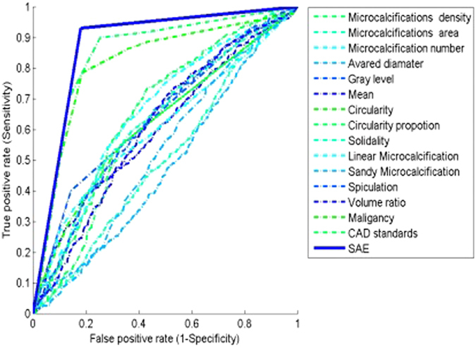
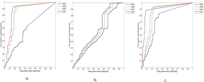
Microcalcification is an effective indicator of early breast cancer. To improve the diagnostic accuracy of microcalcifications, this study evaluates the performance of deep learning-based models on large datasets for its discrimination. A semi-automated segmentation method was used to characterize all microcalcifications. A discrimination classifier model was constructed to assess the accuracies of microcalcifications and breast masses, either in isolation or combination, for classifying breast lesions. Performances were compared to benchmark models. Our deep learning model achieved a discriminative accuracy of 87.3% if microcalcifications were characterized alone, compared to 85.8% with a support vector machine. The accuracies were 61.3% for both methods with masses alone and improved to 89.7% and 85.8% after the combined analysis with microcalcifications. Image segmentation with our deep learning model yielded 15, 26 and 41 features for the three scenarios, respectively. Overall, deep learning based on large datasets was superior to standard methods for the discrimination of microcalcifications. Accuracy was increased by adopting a combinatorial approach to detect microcalcifications and masses simultaneously. This may have clinical value for early detection and treatment of breast cancer.
Figures
References
-
- Cady B. & Chung M. Mammographic screening: no longer controversial. American Journal of Clin Oncol 28(1), 1–4 (2005). - PubMed
-
- American College of Radiology (ACR). Breast imaging reporting and data system (BI-RADS), breast imaging atlas. 4th ed., Reston,Va, Am College Radiology, 1–259 (2003).
-
- Winchester D. P., Jeske J. M. & Goldschmidt R. A. The diagnosis and management of ductal carcinoma in situ of the breast. Am Cancer J Clin 50(3), 184 (2000). - PubMed
-
- Schreer I. & Luttges J. Breast cancer: early detection. Eur J Radiol 11 (Suppl 2), S307–S314 (2001).
Publication types
MeSH terms
LinkOut - more resources
Full Text Sources
Other Literature Sources
Medical
