

Function-structure connectivity in patients with severe brain injury as measured by MRI-DWI and FDG-PET
- PMID: 27273334
- PMCID: PMC6867513
- DOI: 10.1002/hbm.23269
Function-structure connectivity in patients with severe brain injury as measured by MRI-DWI and FDG-PET
Abstract
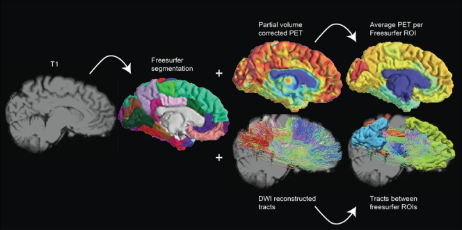
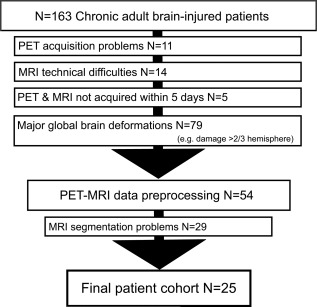
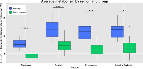
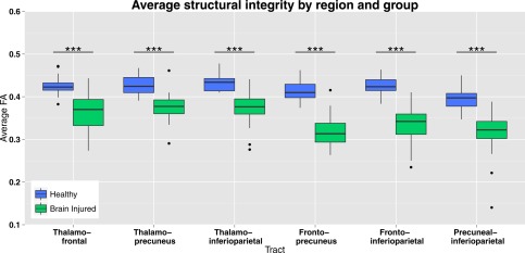
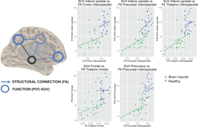
A vast body of literature exists showing functional and structural dysfunction within the brains of patients with disorders of consciousness. However, the function (fluorodeoxyglucose FDG-PET metabolism)-structure (MRI-diffusion-weighted images; DWI) relationship and how it is affected in severely brain injured patients remains ill-defined. FDG-PET and MRI-DWI in 25 severely brain injured patients (19 Disorders of Consciousness of which 7 unresponsive wakefulness syndrome, 12 minimally conscious; 6 emergence from minimally conscious state) and 25 healthy control subjects were acquired here. Default mode network (DMN) function-structure connectivity was assessed by fractional anisotropy (FA) and metabolic standardized uptake value (SUV). As expected, a profound decline in regional metabolism and white matter integrity was found in patients as compared with healthy subjects. Furthermore, a function-structure relationship was present in brain-damaged patients between functional metabolism of inferior-parietal, precuneus, and frontal regions and structural integrity of the frontal-inferiorparietal, precuneus-inferiorparietal, thalamo-inferioparietal, and thalamofrontal tracts. When focusing on patients, a stronger relationship between structural integrity of thalamo-inferiorparietal tracts and thalamic metabolism in patients who have emerged from the minimally conscious state as compared with patients with disorders of consciousness was found. The latter finding was in line with the mesocircuit hypothesis for the emergence of consciousness. The findings showed a positive function-structure relationship within most regions of the DMN. Hum Brain Mapp 37:3707-3720, 2016. © 2016 Wiley Periodicals, Inc.
Keywords: DWI; FDG-PET; default mode network; disorders of consciousness; function-structure coupling.
© 2016 Wiley Periodicals, Inc.
Figures
References
-
- Boly M, Tshibanda L, Vanhaudenhuyse A, Noirhomme Q, Schnakers C, Ledoux D, Boveroux P, Garweg C, Lambermont B, Phillips C (2009): Functional connectivity in the default network during resting state is preserved in a vegetative but not in a brain dead patient. Hum Brain Mapp 30:2393–2400. - PMC - PubMed
-
- Chandra PS, Salamon N, Huang J, Wu JY, Koh S, Vinters HV, Mathern GW (2006): FDG‐PET/MRI coregistration and diffusion‐tensor imaging distinguish epileptogenic tubers and cortex in patients with tuberous sclerosis complex: A preliminary report. Epilepsia 47:1543–1549. - PubMed
-
- De Volder AG, Bol A, Blin J, Robert A, Arno P, Grandin C, Michel C, Veraart C (1997): Brain energy metabolism in early blind subjects: Neural activity in the visual cortex. Brain Res 750:235–244. - PubMed
Publication types
MeSH terms
Substances
LinkOut - more resources
Full Text Sources
Other Literature Sources
