

Review
doi: 10.1038/nsmb.3215.
Atomic-level analysis of membrane-protein structure
Affiliations
- PMID: 27273628
- PMCID: PMC5299386
- DOI: 10.1038/nsmb.3215
Item in Clipboard
Review
Atomic-level analysis of membrane-protein structure
Nat Struct Mol Biol.
.
Abstract
Membrane proteins are substantially more challenging than natively soluble proteins as subjects for structural analysis. Thus, membrane proteins are greatly underrepresented in structural databases. Recently, focused consortium efforts and advances in methodology for protein production, crystallographic analysis and cryo-EM analysis have accelerated the pace of atomic-level structure determination of membrane proteins.
Conflict of interest statement
The author declares no competing financial interests.
Figures
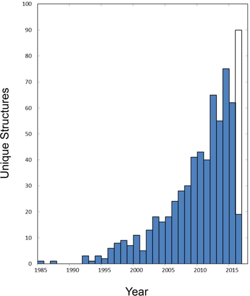
Growth in the production of membrane protein structures. Annual numbers of unique structures are plotted as a function of time. The numbers of unique structures are taken as defined and recorded through 17 March 2016 in the White database of membrane proteins of known structure (http://blanco.biomol.uci.edu/mpstruc/ ). The open portion of the bar for 2016 projects for production continuing at the same rate as for this year to date.

Latticework in a lipidic cubic phase (LCP) crystal of a membrane protein. The crystal structure of TSPO in the apo type 2 LCP lattice is drawn with protein molecules as ribbon diagrams, interstitial monoolein chains in stick representation, and water molecules as red dots.
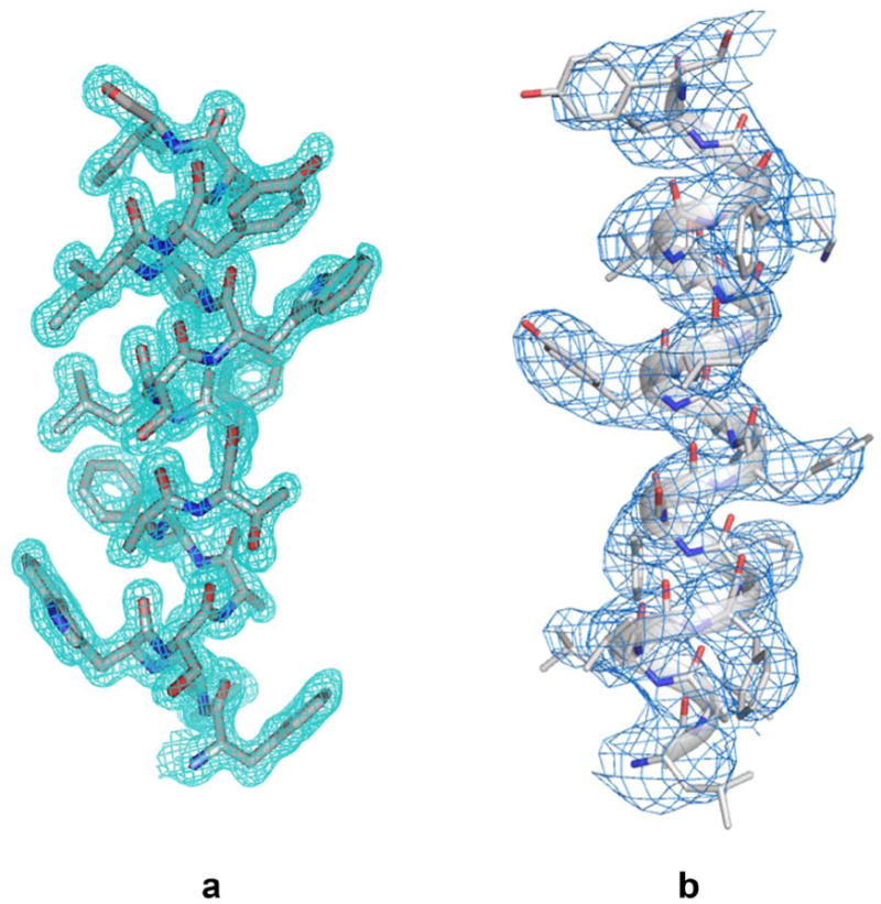
Density distributions for transmembrane helices obtained by different methods. (a) Helix TM5 (residues F136-I49) from TSPO of Bacillus cereus as determined at 1.7 Å resolution from an apo type 2 crystal grown in LCP. Reproduced from Figure S2 of Ref. with permission by Science. (b) Helix TM3 (residues L171-Y189) of presenilin from the cryo-EM structure of human γ-secretase at 3.4 Å resolution. Adapted from Extended Data Figure 2 of Ref. with permission by Nature.
References
-
- Overington JP, Al-Lazikani B, Hopkins AL. How many drug targets are there? Nat Rev Drug Discov. 2006;5:993–996. - PubMed
-
- Henderson R, Unwin PN. Three-dimensional model of purple membrane obtained by electron microscopy. Nature. 1975;257:28–32. - PubMed
-
- Deisenhofer J, Epp O, Miki K, Huber R, Michel H. Structure of the protein subunits in the photosynthetic reaction centre of Rhodopseudomonas viridis at 3Å resolution. Nature. 1985;318:618–624. - PubMed
-
- Doyle DA, et al. The structure of the potassium channel: molecular basis of K+ conduction and selectivity. Science. 1998;280:69–77. - PubMed
Publication types
MeSH terms
Substances
Grants and funding
LinkOut - more resources
Full Text Sources
Other Literature Sources
