. 2016 Jun 28;113(26):7124-9.
doi: 10.1073/pnas.1521738113. Epub 2016 Jun 6.

PROTAC-induced BET protein degradation as a therapy for castration-resistant prostate cancer

Affiliations

PROTAC-induced BET protein degradation as a therapy for castration-resistant prostate cancer

Kanak Raina et al. Proc Natl Acad Sci U S A. .

Abstract

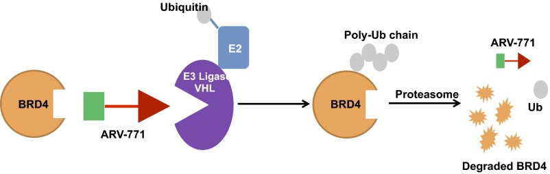
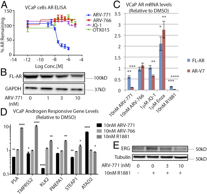
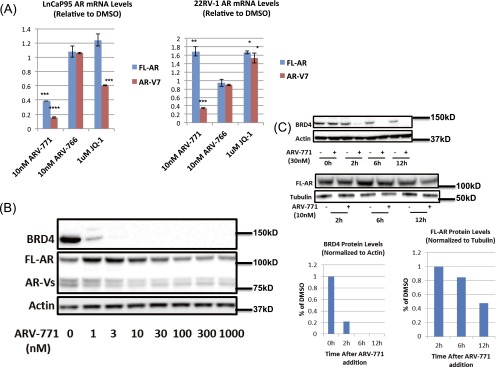
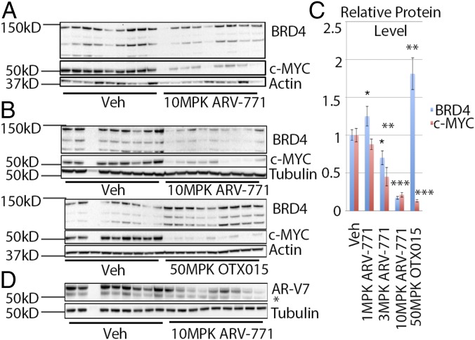
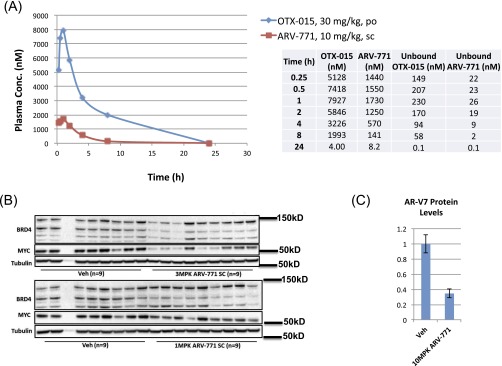
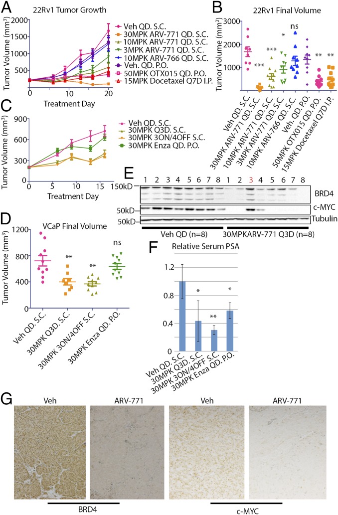
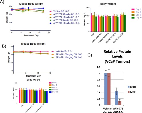
Prostate cancer has the second highest incidence among cancers in men worldwide and is the second leading cause of cancer deaths of men in the United States. Although androgen deprivation can initially lead to remission, the disease often progresses to castration-resistant prostate cancer (CRPC), which is still reliant on androgen receptor (AR) signaling and is associated with a poor prognosis. Some success against CRPC has been achieved by drugs that target AR signaling, but secondary resistance invariably emerges, and new therapies are urgently needed. Recently, inhibitors of bromodomain and extra-terminal (BET) family proteins have shown growth-inhibitory activity in preclinical models of CRPC. Here, we demonstrate that ARV-771, a small-molecule pan-BET degrader based on proteolysis-targeting chimera (PROTAC) technology, demonstrates dramatically improved efficacy in cellular models of CRPC as compared with BET inhibition. Unlike BET inhibitors, ARV-771 results in suppression of both AR signaling and AR levels and leads to tumor regression in a CRPC mouse xenograft model. This study is, to our knowledge, the first to demonstrate efficacy with a small-molecule BET degrader in a solid-tumor malignancy and potentially represents an important therapeutic advance in the treatment of CRPC.

Keywords: BET; BRD4; PROTAC; prostate; protein degradation.


Conflict of interest statement

Conflict of interest statement: C.M.C. is the founder and Chief Scientific Advisor of, and possesses shares in, Arvinas, LLC.

Figures

Fig. S1.
Fig. S1.
BRD4 PROTAC schematic.
Fig. 1.
Fig. 1.
ARV-771 is a potent pan-BET degrader. (A) Chemical structures of ARV-771 and the inactive diastereomer ARV-766, which is unable to bind VHL. (B) Incubation of the indicated CRPC cell lines with ARV-771 for 16 h results in depletion of BRD2/3/4 in 22Rv1, VCaP, and LnCaP95 cells. The Western blot is representative of three independent experiments (n = 3). (C) ARV-771 treatment for 16 h results in suppression of cellular c-MYC levels measured by ELISA. The assay was performed in triplicate (n = 3). (D) ARV-771–mediated c-MYC suppression occurs at the mRNA level, as determined by qPCR analysis following 16-h treatment at the indicated concentrations. c-MYC levels were also monitored in the same cell lines by qPCR following a 16-h treatment with either 1 μM ARV-766 or 1 μM OTX015. The results shown represent an average of two biological replicates, each measured in triplicate (n = 3). (E) ARV-771–mediated BRD4 degradation at 8 h is blocked by 30-min pretreatment with either an excess of VHL ligand ARV-056 (10 μM) or the proteasome inhibitor carfilzomib (1 μM). The Western blot is representative of two independent experiments (n = 2). All data represent mean values ± SEM (*P < 0.05; **P < 0.01; ***P < 0.001; ****P < 0.0001). P values were determined using GraphPad Prism using an unpaired parametric t test with Welch’s correction.
Fig. S2.
Fig. S2.
(A) Kd values of ARV-771 and ARV-766 against BRD1 and BRD2. (B) VHL ligand ARV-056. (C) BRD4 immunoprecipitated from 22Rv1 cells transfected with HA-Ubiquitin. (D) RNAi confirmation of BRD4 band identity in vitro. (E) ARV-771 treatment results in a change in 22Rv1 cellular morphology consistent with apoptosis. (Magnification: 20×.)
Fig. 2.
Fig. 2.
ARV-771 treatment results in cell death in CRPC cell lines. (A) Antiproliferative effect of ARV-771 in CRPC cell lines after 72-h treatment. Experiments were performed in triplicate (n = 3). (B) Treatment with ARV-771 for 24 h leads to caspase activation in CRPC cell lines. Experiments were performed in triplicate (n = 3). (C) Quantification of results in A. (D) Quantification of results in B. (E) A 24-h ARV-771 treatment leads to PARP cleavage in 22Rv1 cells. All data represent mean values ± SEM (*P < 0.05; **P < 0.01; ***P < 0.001; ****P < 0.0001). P values were determined using GraphPad Prism using an unpaired t test with a false-discovery rate of 1%.
Fig. 3.
Fig. 3.
ARV-771 treatment attenuates AR signaling. (A) ARV-771 treatment for 16 h results in lower FL-AR levels as measured by ELISA. The assay was performed in triplicate (n = 3). (B) ARV-771–mediated lowering of FL-AR levels demonstrated by immunoblotting after a 16-h treatment. The Western blot is representative of two independent experiments (n = 2). (C) mRNA levels of FL-AR and AR-V7 are lowered by a 16-h ARV-771 treatment in VCaP cells, as determined by qPCR analysis. The result shown is an average of two biological replicates, each measured in triplicate (n = 3). (D) Levels of androgen-responsive genes in response to a 16-h ARV-771 treatment. The result is an average of two biological replicates, each measured in triplicate (n = 3). (E) ERG induction by a 16-h treatment with R1881 in VCaP cells is blocked by a 1-h ARV-771 pretreatment. All data represent mean values ± SEM (*P < 0.05; **P < 0.01; ***P < 0.001; ****P < 0.0001). P values were determined using GraphPad Prism using an unpaired parametric t test with Welch’s correction.
Fig. S3.
Fig. S3.
(A) FL-AR and AR-V7 mRNA levels in LnCaP95 and 22Rv1 cells. (B) AR protein levels in 22Rv1 cells in response to ARV-771. (C) Kinetics of depletion of BRD4 and FL-AR following ARV-771 treatment in VCaP cells. *P < 0.05; **P < 0.01; ***P < 0.001; ****P < 0.0001.
Fig. 4.
Fig. 4.
ARV-771 is a potent in vivo PROTAC. (A) BRD4 down-regulation and c-MYC suppression in 22Rv1 tumor xenografts implanted in Nu/Nu mice after daily subcutaneous administration of 10 mg/kg ARV-771 for 3 d. Each treatment cohort contained nine animals (n = 9). (B) Effect of ARV-771 and OTX015 on BRD4 and c-MYC levels by immunoblotting in a 14-d 22Rv1 tumor xenograft study. Each treatment cohort contained nine animals (n = 9). (C) Quantification of results in B and Fig. S4B. Quantification of the highest BRD4 band in B is shown. Quantification of all three bands gave the same result. (D) AR-V7 levels in 22Rv1 tumor xenografts are also lowered by daily subcutaneous injection of 10 mg/kg ARV-771 in a 14-d 22Rv1 tumor xenograft study. All data represent mean values ± SEM. In all experiments, tumors and plasma were harvested 8 h after the last dose for analysis (*P < 0.05; **P < 0.01; ***P < 0.001; ****P < 0.0001). P values were determined using GraphPad Prism using an unpaired parametric t test with Welch’s correction.
Fig. S4.
Fig. S4.
(A) PK profile of ARV-771 and OTX015 in mice after a single dose with the indicated route and concentration. (B) Down-regulation of BRD4 and MYC with 3 mg/kg and 10 mg/kg ARV-771 in a 14-d 22Rv1 tumor xenograft study. (C) Quantification of the Western blot in Fig. 4D.
Fig. 5.
Fig. 5.
ARV-771 is efficacious in multiple tumor xenograft models of CRPC. (A) Results of an efficacy study in 22Rv1 tumor xenografts implanted in Nu/Nu mice showing tumor regression with 30 mg/kg subcutaneously once daily ARV-771 dosing. Each treatment cohort contained 10 animals (n = 10). (B) Scatter plot of the results from A demonstrating dose-dependent TGI with ARV-771. (C) Intermittent dosing schedules of ARV-771 are sufficient to induce TGI in CB17 SCID mice bearing VCaP tumor xenografts. Each treatment cohort contained 10 animals (n = 10). (D) Scatter plot of the results from C. (E) ARV-771 dosing results in pharmacodynamic depletion of BRD4 and suppression of c-MYC in the VCaP tumor xenograft model. (F) PSA levels in serum from the mice in C were analyzed by ELISA, showing suppression of levels with ARV-771 treatment. The numbers of replicates for treatments shown in the graph were 8, 5, 8, and 8, respectively. (G) Immunohistochemical staining of tumor samples from vehicle- and ARV-771–treated VCaP tumor-bearing mice. All data represent mean values ± SEM. In all experiments, tumors and plasma were harvested 8 h after the last dose (*P < 0.05; **P < 0.01; ***P < 0.001; ****P < 0.0001). P values were determined using GraphPad Prism using an unpaired parametric t test with Welch’s correction.
Fig. S5.
Fig. S5.
(A) Body weights of mice in the 22Rv1 xenograft study. (B) Body weights of mice in the VCaP xenograft study. (C) Quantification of immunoblots in Fig. 5E.
Fig. S6.
Fig. S6.
Synthetic scheme for ARV-771 and ARV-766.

References

    1. Wong YNS, Ferraldeschi R, Attard G, de Bono J. Evolution of androgen receptor targeted therapy for advanced prostate cancer. Nat Rev Clin Oncol. 2014;11(6):365–376. - PubMed
    1. Aragon-Ching JB. The evolution of prostate cancer therapy: Targeting the androgen receptor. Front Oncol. 2014;4:295. - PMC - PubMed
    1. Trewartha D, Carter K. Advances in prostate cancer treatment. Nat Rev Drug Discov. 2013;12(11):823–824. - PubMed
    1. Mottet N, et al. EAU guidelines on prostate cancer. Part II: Treatment of advanced, relapsing, and castration-resistant prostate cancer. Eur Urol. 2011;59(4):572–583. - PubMed
    1. Chen CD, et al. Molecular determinants of resistance to antiandrogen therapy. Nat Med. 2004;10(1):33–39. - PubMed

Publication types

MeSH terms