

Case Reports
doi: 10.1016/j.jdcr.2016.03.010.
eCollection 2016 May.
Keratoacanthoma centrifugum marginatum: A diagnostic and therapeutic challenge
Affiliations
- PMID: 27274538
- PMCID: PMC4885138
- DOI: 10.1016/j.jdcr.2016.03.010
Item in Clipboard
Case Reports
Keratoacanthoma centrifugum marginatum: A diagnostic and therapeutic challenge
JAAD Case Rep.
.
No abstract available
Keywords: 5-fluorouracil; KA, keratoacanthoma; KCM, keratoacanthoma centrifugum marginatum; dermatopathology; intralesional; keratoacanthoma; keratoacanthoma centrifugum marginatum; trauma.
Figures
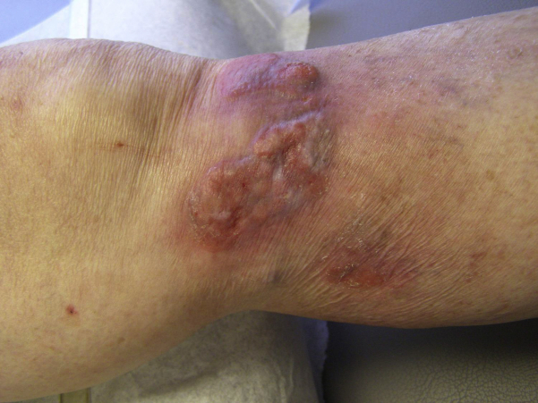
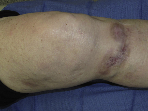
Keratoacanthoma centrifugum marginatum. The lesion began as a single erythematous papule that progressed to an irregular 7.5- × 3.5-cm boggy tumor on the left anterior knee approximately 1 year after localized trauma.
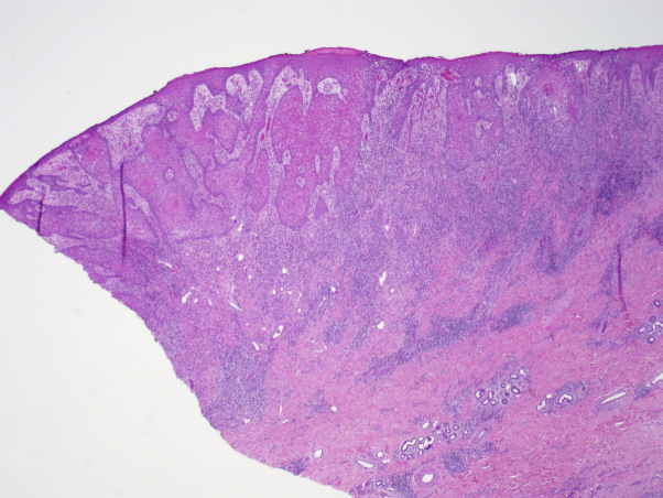
A low-power view of the incisional biopsy with zonal changes from the periphery to the center of the lesion. The outer edge is characterized by pseudoepitheliomatous hyperplasia and a dense mixed inflammatory infiltrate both superficially as well as deep. Centrally, the lesion shows fibrosis and a lack of epidermal hyperplasia. (Hematoxylin-eosin stain; original magnification: ×2.)
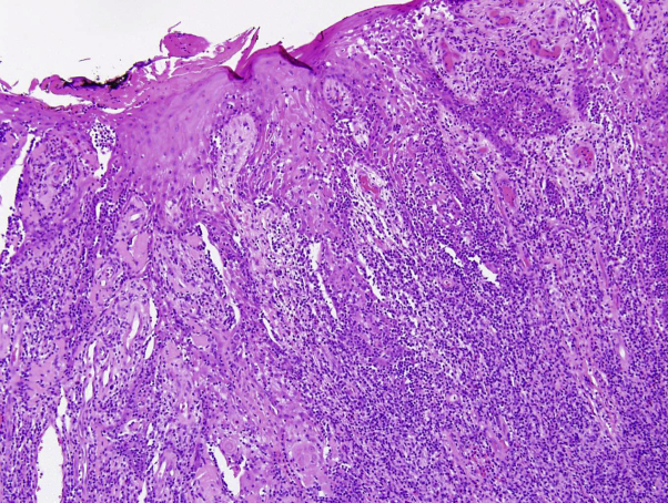
A high-power view shows islands of keratinocytes with squamous pearls and minimal atypia underlying pseudoepitheliomatous hyperplasia. (Hematoxylin-eosin stain; original magnification: ×20.)
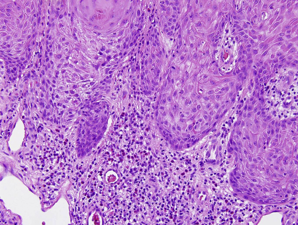
A high-power view shows the eosinophilic, glassy cytoplasm of the keratinocytes, minimal atypia, and a single mitotic figure near the basal layer. The dermal inflammatory infiltrate is a mixture of lymphocytes, histiocytes, eosinophils, and a few plasma cells. (Hematoxylin-eosin stain; original magnification: ×40.)
Clinical photo shows complete response within weeks of receiving 2 courses of intralesional 5-fluorouracil injections.
References
-
- V'Ickova-Laskoska M.T., Laskoski D.S. “Keratoacanthoma centrifugum marginatum: a rare atypical variant of keratoacanthoma”. Clin Exp Dermatol. 2007;33:259–261. - PubMed
-
- Mangas C., Bielsa I., Ribera M., Fernandez-Fiqueras M.T., Ferrandiz C. “A Case of Multiple Keratoacanthoma Centrifugum Marginatum”. Dermatol Surg. 2004;30:803–806. - PubMed
-
- Miedzinski F., Kozakiewicz J. “Das keratoacanthoma centrifugum eine besandera varietat des keratoacanthomas”. Hautarzt. 1962;13:348–352. - PubMed
Publication types
LinkOut - more resources
Full Text Sources
Other Literature Sources