Review
. 2016:2016:6810562.
doi: 10.1155/2016/6810562. Epub 2016 May 4.

Biomaterial Applications in Cell-Based Therapy in Experimental Stroke

Affiliations
Review

Biomaterial Applications in Cell-Based Therapy in Experimental Stroke

Ligia S B Boisserand et al. Stem Cells Int. 2016.

Abstract

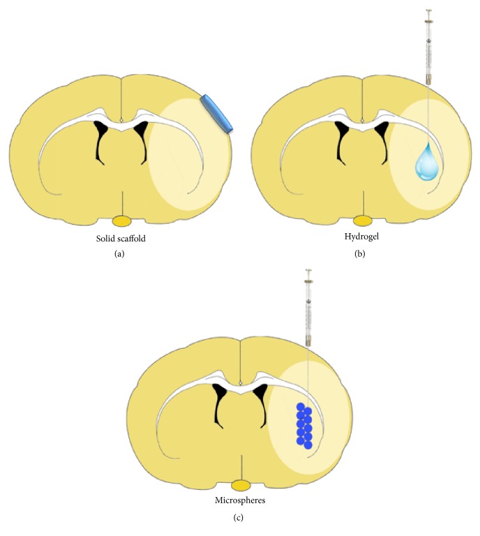
Stroke is an important health issue corresponding to the second cause of mortality and first cause of severe disability with no effective treatments after the first hours of onset. Regenerative approaches such as cell therapy provide an increase in endogenous brain structural plasticity but they are not enough to promote a complete recovery. Tissue engineering has recently aroused a major interesting development of biomaterials for use into the central nervous system. Many biomaterials have been engineered based on natural compounds, synthetic compounds, or a mix of both with the aim of providing polymers with specific properties. The mechanical properties of biomaterials can be exquisitely regulated forming polymers with different stiffness, modifiable physical state that polymerizes in situ, or small particles encapsulating cells or growth factors. The choice of biomaterial compounds should be adapted for the different applications, structure target, and delay of administration. Biocompatibilities with embedded cells and with the host tissue and biodegradation rate must be considerate. In this paper, we review the different applications of biomaterials combined with cell therapy in ischemic stroke and we explore specific features such as choice of biomaterial compounds and physical and mechanical properties concerning the recent studies in experimental stroke.


Figures

Figure 1
Figure 1
Schematic illustration of different biomaterial applications on ischemic brain. (a) Solid brain scaffolds for surface application and gradual liberation of cells, drugs, or growth factors. (b) Injectable hydrogel, in liquid phase with an in situ gelation. (c) Microspheres for gradual intracerebral delivery.
Figure 2
Figure 2
Different experimental steps for intracerebral graft of cell-biomaterial after stroke. Scale bar = 100 μm. (a) In vitro biocompatibility: after mixing human mesenchymal stem cells (MSC) within hyaluronic acid (HA) hydrogel (Hystem HP, Sigma: hyaluronan+polyethylene glycol diacrylate), MSC survived into the gel during several days in culture (A1) without cell death (A2, propidium iodide cell dead assay). Cell survival and spreading into the HA gel were assessed in one-week culture (A3) using confocal microscopy and confocal microscopy stacks and viable cell labelling (A4 and A5, Cell Tracker Green CMFDA, Life). (b) Intracerebral transplantation: one week after experimental ischemic stroke in rat, magnetic resonance imaging was used to determine the injection site into the stroke cavity near plastic areas surrounding the lesion (B1). Coordinates for stereotactic injection were defined using anatomic atlas (Watson-Paxinos) (B2). By histology, the stereotactic tract can be macroscopically observed (B3, crysostat section). (c) In vivo biocompatibility and effects: ex vivo brain immunohistology demonstrated cell survival into the graft site such as human MSC identification in stroke lesion (C1, human-specific monoclonal antibody to nuclear antigen, MAB1281, 1/1,000, Chemicon) without cell migration in contralateral hemisphere (C2). Additional experiments must be done to assess long-term cell differentiations and host integration, hydrogel biodegradation, local inflammatory response, and behavior recovery effects.
Figure 3
Figure 3
Representative images of in vivo and ex vivo detection of hyaluronan-acid (HA) hydrogel. (a)Magnetic Resonance Imaging (MRI) weighed in T2, hydrogel detected at different time points (days one, seven, and fourteen after administration). (b) Cresyl violet staining of HA hydrogel acquired two weeks after administration, noted in (×2) (b) and (c) (x10) magnification. These images demonstrate efficient local gel formation instead of liquid diffusion which would be due to a delayed polymerization after infusion and the in vivo stability of HA hydrogel.

References

    1. Beal C. C. Gender and stroke symptoms: a review of the current literature. The Journal of Neuroscience Nursing. 2010;42(2):80–87. doi: 10.1097/jnn.0b013e3181ce5c70. - DOI - PubMed
    1. Wang H.-R., Chen M., Wang F.-L., et al. Comparison of therapeutic effect of recombinant tissue plasminogen activator by treatment time after onset of acute ischemic stroke. Scientific Reports. 2015;5 doi: 10.1038/srep11743.11743 - DOI - PMC - PubMed
    1. Duncan P. W., Zorowitz R., Bates B., et al. Management of adult stroke rehabilitation care: a clinical practice guideline. Stroke. 2005;36(9):e100–e143. doi: 10.1161/01.str.0000180861.54180.ff. - DOI - PubMed
    1. Detante O., Jaillard A., Moisan A., et al. Biotherapies in stroke. Revue Neurologique. 2014;170(12):779–798. doi: 10.1016/j.neurol.2014.10.005. - DOI - PubMed
    1. Pellegrini L., Bennis Y., Guillet B., Velly L., Bruder N., Pisano P. La thérapie cellulaire de l’accident vasculaire cérébral ischémique: du mythe à la réalité. Revue Neurologique. 2013;169(4):291–306. doi: 10.1016/j.neurol.2012.08.009. - DOI - PubMed