. 2015 Mar 15;3(1):99-104.
doi: 10.3889/oamjms.2015.016. Epub 2015 Jan 22.

Achieving Higher Diagnostic Results in Stereotactic Brain Biopsy by Simple and Novel Technique

Affiliations

Achieving Higher Diagnostic Results in Stereotactic Brain Biopsy by Simple and Novel Technique

Salih Gulsen. Open Access Maced J Med Sci. .

Abstract

Background: Neurosurgeons have preferred to perform the stereotactic biopsy for pathologic diagnosis when the intracranial pathology located eloquent areas and deep sites of the brain.

Aim: To get a higher ratio of definite pathologic diagnosis during stereotactic biopsy and develop practical method.

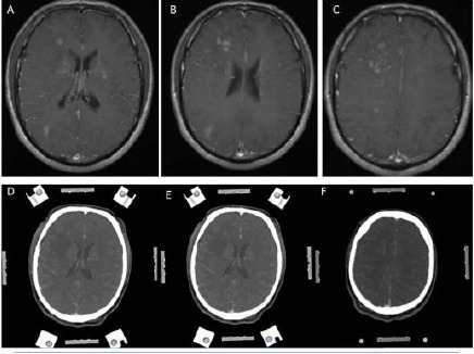
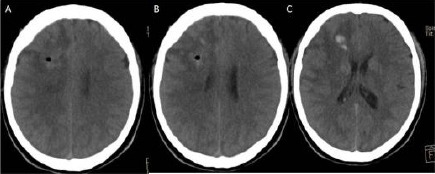
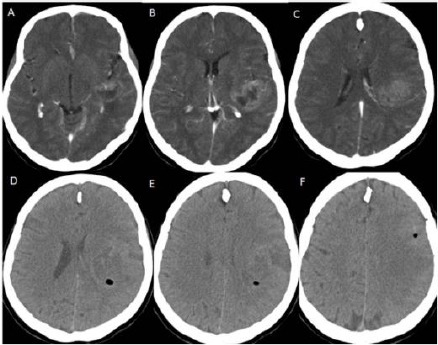
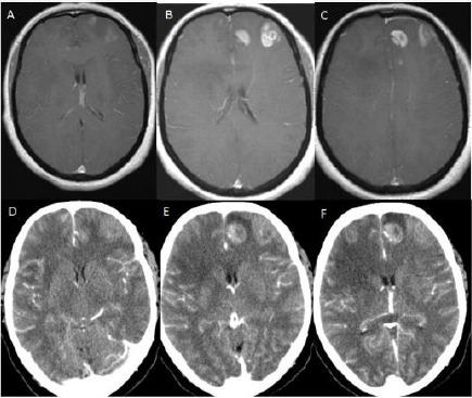
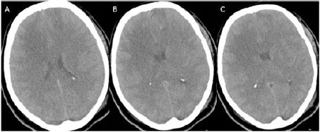
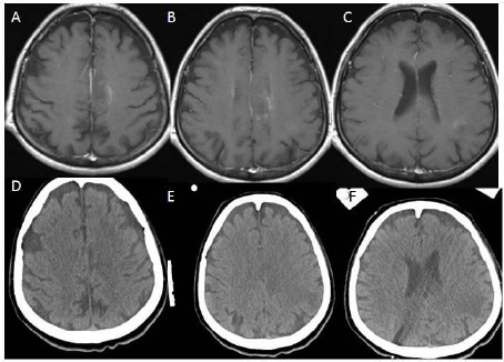
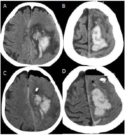
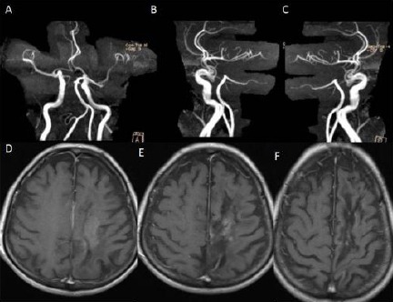
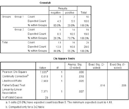
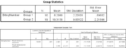
Material and methods: We determined at least two different target points and two different trajectories to take brain biopsy during stereotactic biopsy. It is a different way from the conventional stereotactic biopsy method in which one point has been selected to take a biopsy. We separated our patients into two groups, group 1 (N=10), and group 2 (N= 19). We chose one target to take a biopsy in group 1, and two different targets and two different trajectories in group 2. In group 2, one patient underwent craniotomy due to hemorrhage at the site of the biopsy during tissue biting. However, none of the patients in both groups suffered any neurological complication related biopsy procedure.

Results: In group 1, two of 10 cases, and, in group 2, fourteen of 19 cases had positive biopsy harvesting. These results showed statistically significant difference between group 1 and group 2 (P<0.05).

Conclusions: Regarding these results, choosing more than one trajectories and taking at least six specimens from each target provides higher diagnostic rate in stereotaxic biopsy taking method.

Keywords: Brain Tumour; Diagnostic rate; Diffuse infilitrative; Eloquent Areas; Stereotactic Biopsy.


Figures

Figure 1
Figure 1
(Group 2, case 11): A, B, C - Contrast-enhanced (Gadolinium) T1 weighted cranial MRI in axial sections showing diffuse infiltrative multiple lesions around lateral ventricles and centrum semiovale. D, E, F - Contrast-enhanced (Metrizamide) cranial CT in axial sections showing weak contrast enhancement, and its contrast enhancing regions is corresponding with his MRI appearances. Note: This cranial CT was taken with a stereotactic frame before the operation.
Figure 2
Figure 2
A, B, C: Cranial CT following the procedure showing little amount of hemorrhage that is not caused any symptoms and signs.
Figure 3
Figure 3
(Group 2, case 8): A, B, C - Contrast-enhanced (Metrizamide) cranial CT in axial sections showing weak contrast enhancement at the insular region. Note: This cranial CT was taken with a stereotactic frame before the operation. D, E, F - Cranial CT following the procedure showing no hemorrhage. Also, this cranial CT showing two different target points with two different trajectories marked oval shaped hypodens areas at the centrum semiovale and around the insula on the left hemisphere.
Figure 4
Figure 4
(Group 2, Case 2): A, B, C - Contrast-enhanced (Gadolinium) T1 weighted cranial MRI in axial sections showing multiple hyperintense lesions at the anterior part of the frontal lobe of the left hemisphere. D, E, F - Contrast-enhanced (Metrizamide) cranial CT in axial sections showing weak contrast enhancement at the anterior part of the frontal lobe, and its contrast enhancing region is corresponding with his MRI appearances. Note: This cranial CT was taken with a stereotactic frame before the operation.
Figure 5
Figure 5
A, B, C: Cranial CT in axial sections following the biopsy procedure showing very little amount of bleeding.
Figure 6
Figure 6
(Group 2, case 19): A, B, C - Contrast-enhanced (Gadolinium) T1 weighted cranial MRI in axial sections showing weak hyperintense lesions on the left parasagittal region of the centrum semiovale and around left lateral ventricular area. D, E, F - Cranial CT without contrast in axial sections showing hypodense areas on the left parasagittal region of the centrum semiovale and around left lateral ventricular region corresponding with hypodense areas of cranial MRI.
Figure 7
Figure 7
A, B: Cranial CT of the axial sections showing hematoma at the left centrum semiovale after biopsy procedure; C, D: After hematoma evacuation.
Figure 8
Figure 8
A, B, C - Cranial MR angiography showing no damage any arterial structures due to the biopsy procedure. D, E, F - Cranial MRI showing complete resorption of the hematoma 7 months later of the operation.
Figure 9
Figure 9
Chi-Square test is showing statistically significant difference between group 1 and group 2 regarding histopathological results of the cases.
Figure 10
Figure 10
Independent sample Student T test is showing statistically significant difference between group 1 and group 2 concerning the number of taken biopsy.

References

    1. Apuzzo ML, Sabshin JK. Computed tomographic guidance stereotaxis in the management of intracranial mass lesions. Neurosurgery. 1983;12:277–85. - PubMed
    1. Ostertag CB, Mennel HD, Kiessling M. Stereotactic biopsy of brain tumors. Surg Neurol. 1980;14:275–83. - PubMed
    1. Edner G. Stereotactic biopsy of intracranial space occupying lesions. Acta Neurochir (Wien) 1981;57:213–34. - PubMed
    1. Grunert P, Ungersbock K, Bohl J, Kitz K, Hopf N. Results of 200 intracranial stereotactic biopsies. Neurosurgery. 1994;17:59–66. - PubMed
    1. Hall WA. The safety and efficacy of stereotactic biopsy for intracranial lesions. Cancer. 1998;82:1749–55. - PubMed

LinkOut - more resources