Review
. 2016 Jun;10(3):189-99.
doi: 10.1007/s11571-016-9379-z. Epub 2016 Feb 3.

Towards a fourth spatial dimension of brain activity

Affiliations
Review

Towards a fourth spatial dimension of brain activity

Arturo Tozzi et al. Cogn Neurodyn. 2016 Jun.

Abstract

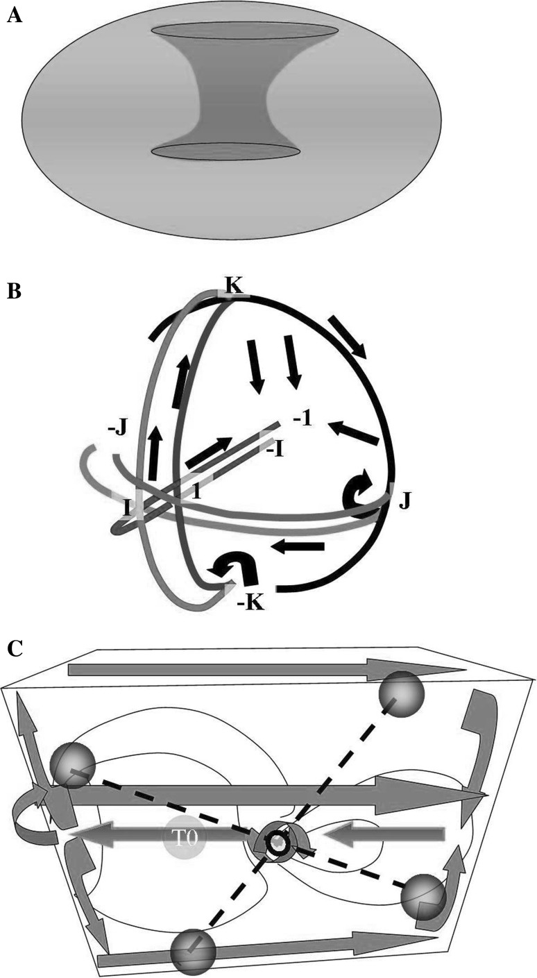
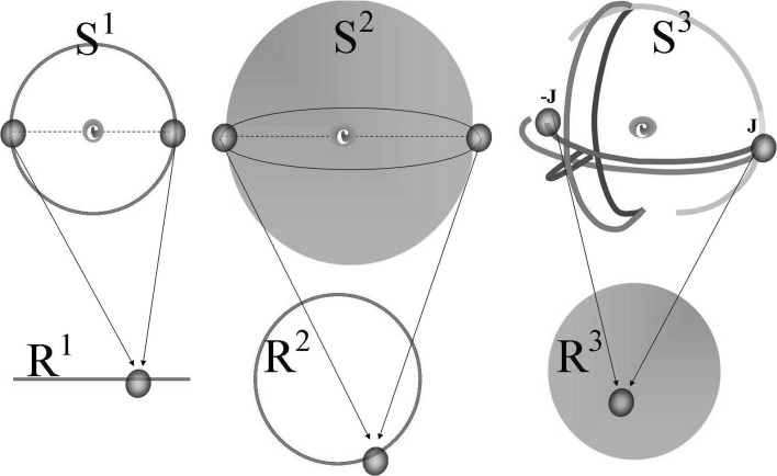
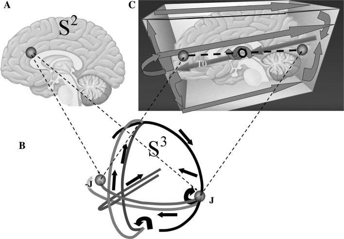
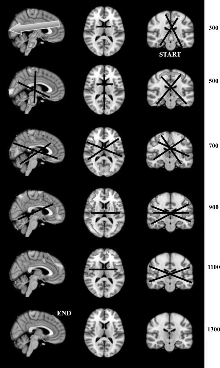
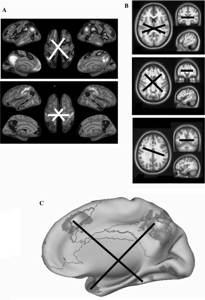
Current advances in neurosciences deal with the functional architecture of the central nervous system, paving the way for general theories that improve our understanding of brain activity. From topology, a strong concept comes into play in understanding brain functions, namely, the 4D space of a "hypersphere's torus", undetectable by observers living in a 3D world. The torus may be compared with a video game with biplanes in aerial combat: when a biplane flies off one edge of gaming display, it does not crash but rather it comes back from the opposite edge of the screen. Our thoughts exhibit similar behaviour, i.e. the unique ability to connect past, present and future events in a single, coherent picture as if we were allowed to watch the three screens of past-present-future "glued" together in a mental kaleidoscope. Here we hypothesize that brain functions are embedded in a imperceptible fourth spatial dimension and propose a method to empirically assess its presence. Neuroimaging fMRI series can be evaluated, looking for the topological hallmark of the presence of a fourth dimension. Indeed, there is a typical feature which reveal the existence of a functional hypersphere: the simultaneous activation of areas opposite each other on the 3D cortical surface. Our suggestion-substantiated by recent findings-that brain activity takes place on a closed, donut-like trajectory helps to solve long-standing mysteries concerning our psychological activities, such as mind-wandering, memory retrieval, consciousness and dreaming state.

Keywords: Borsuk-Ulam theorem; Brain; Central nervous system; Fourth dimension; Hypersphere; Manifold.


Figures

Fig. 1
Fig. 1
Different ways to depict a hypersphere. a How two 2-spheres glued together along their spherical boundary give rise to a donut-shaped Clifford torus. b Another way to depict a hypersphere: the superimposition of two 2-spheres (which circumferences are glued together) gives rise to a glome. Some of the quaternion rotations are depicted by the straight and curved arrows. c 3D projection of a hypersphere. The lines on the left enlarge in diameter, forming a circle of increasing circumference on the left surface of the 3D space. Conversely, on the opposite right side, the lines shrink and give rise to a circle of decreasing circumference on the right surface of the 3D space. See text for further details. The dotted lines and the black spheres depict some of the possible antipodal points predicted by the Borsuk Ulam Theorem (to give another example, J and −J are antipodal points in a)
Fig. 2
Fig. 2
The Borsuk-Ulam theorem for different values of Sn. S1 depicts a circumference, S2 a common sphere, S3 a hypersphere, while R1 portrays a line, R2 a circumference and R3 a common sphere. Note that the two antipodal points in every sphere Sn project to a single point in the corresponding space Rn, and vice versa
Fig. 3
Fig. 3
The concept of hypersphere in the framework of brain functional activity. According to the Borsuk-Ulam theorem, the activation of a single point on the S2 brain surface (Fig. 1a) leads to the activation of two antipodal points (corresponding in this case to the quaternionic points J and −J) on the S3 brain surface (b). The simultaneous activation of the S3 antipodal points displayed in b can be also evaluated on the 3D cortical surface (c), provided the brain is embedded in a 3D space containing the 4D Clifford torus (the same 3D space described in Fig. 1c). The nomenclature is borrowed from c
Fig. 4
Fig. 4
Video frames, modified from Mitra et al. (2015), showing lag threads computed from real BOLD resting state rs-fMRI data in a group of 688 subjects, obtained from the Harvard-MGH Brain Genomics Superstruct Project (see Mitra et al. for further technical details). Note the widely diffused presence of BUT hallmarks (black lines) at different times and in different brain projections
Fig. 5
Fig. 5
Examples from functional neuroimaging real data showing how the predicted antipodal points can be correctly identified: given one point (a brain signal), there is a second point (another brain signal) at the opposite end of a straight line segment connecting them and passing for the center (white or black lines). a Decomposition of Spontaneous Brain Activity into Distinct fMRI Co-activation Patterns (Liu et al. 2013). b Different clusters during resting-state fMRI scanning, evaluated through innovation-driven co-activation patterns (called iCAPs) (Karahanoglu and Van De Ville 2015). c Significant meta-analytic clusters of fMRI temporal activation associated with mind-wandering and related spontaneous thought processes (Fox et al. 2015)

References

    1. Afraimovich V, Tristan I, Varona P, Rabinovich M. Transient dynamics in complex systems: heteroclinic sequences with multidimensional unstable manifolds. Discontin Nonlinearity Complex. 2013;2(1):21–41. doi: 10.5890/DNC.2012.11.001. - DOI
    1. Ajilore O, et al. Constructing the resting state structural connectome. Front Neuroinform. 2013;7:30. doi: 10.3389/fninf.2013.00030. - DOI - PMC - PubMed
    1. Amft M, et al. Definition and characterization of an extended social-affective default network. Brain Struct Funct. 2015;220:1031–1049. doi: 10.1007/s00429-013-0698-0. - DOI - PMC - PubMed
    1. Andrews-Hanna JR, et al. The default network and self-generated thought: component processes, dynamic control, and clinical relevance. Ann NY Acad Sci. 2014;1316:29–52. doi: 10.1111/nyas.12360. - DOI - PMC - PubMed
    1. Barttfeld P, et al. Signature of consciousness in the dynamics of resting-state brain activity. Proc Natl Acad Sci USA. 2015;112:887–892. doi: 10.1073/pnas.1418031112. - DOI - PMC - PubMed

LinkOut - more resources