. 2016 Mar 7:2:16017.
doi: 10.1038/cddiscovery.2016.17.

Loss of function of Ywhah in mice induces deafness and cochlear outer hair cells' degeneration

Affiliations

Loss of function of Ywhah in mice induces deafness and cochlear outer hair cells' degeneration

L Buret et al. Cell Death Discov. .

Abstract

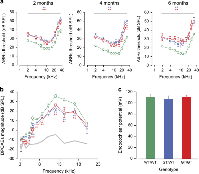
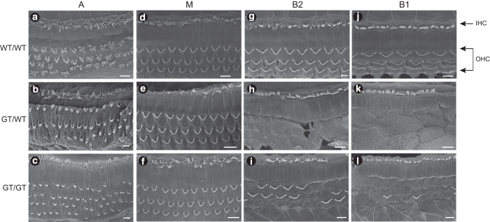
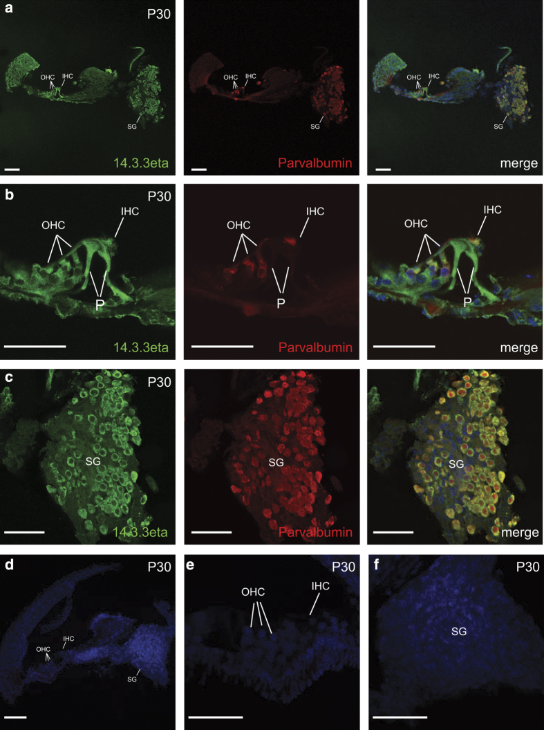
In vertebrates, 14-3-3 proteins form a family of seven highly conserved isoforms with chaperone activity, which bind phosphorylated substrates mostly involved in regulatory and checkpoint pathways. 14-3-3 proteins are the most abundant protein in the brain and are abundantly found in the cerebrospinal fluid in neurodegenerative diseases, suggesting a critical role in neuron physiology and death. Here we show that 14-3-3eta-deficient mice displayed auditory impairment accompanied by cochlear hair cells' degeneration. We show that 14-3-3eta is highly expressed in the outer and inner hair cells, spiral ganglion neurons of cochlea and retinal ganglion cells. Screening of YWHAH, the gene encoding the 14-3-3eta isoform, in non-syndromic and syndromic deafness, revealed seven non-synonymous variants never reported before. Among them, two were predicted to be damaging in families with syndromic deafness. In vitro, variants of YWHAH induce mild mitochondrial fragmentation and severe susceptibility to apoptosis, in agreement with a reduced capacity of mutated 14-3-3eta to bind the pro-apoptotic Bad protein. This study demonstrates that YWHAH variants can have a substantial effect on 14-3-3eta function and that 14-3-3eta could be a critical factor in the survival of outer hair cells.


Figures

Figure 1
Figure 1
Invalidation of Ywhah induces a mild deafness but does not affect the functioning of the stria vascularis. (a) Audiograms established in 14-3-3etaWT/WT (n=18), 14-3-3etaGT/WT (n=16) and 14-3-3etaGT/GT (n=20) by measuring the brainstem evoked potential thresholds at 10 frequencies ranging from 2 to 32 kHz. Data show a mild (10–20 decibels (dB)) but very significant (P<0.01 for all tested frequencies) threshold elevation in 14-3-3etaGT/WT (blue triangles) and 14-3-3etaGT/GT (red squares) mice as compared with 14-3-3etaWT/WT (green circles) at 2, 4 and 6 months. (b) This panel displays cubic DPOAE (2f 1f 2) audiograms represented as a function of f 2. The magnitude of the distortion product is slightly reduced in 14-3-3etaGT/WT (blue triangles; n=14) and 14-3-3etaGT/GT (red squares; n= 6) as compared with 14-3-3etaWT/WT mice (green circles; n=14). This reduction is significant between 6 and 20 kHz. The gray line represents the noise floor. (c) Endocochlear potentials were recorded in 14-3-3etaWT/WT (green bar; n=5), 14-3-3etaGT/WT (blue bar; n=5) and 14-3-3etaGT/GT mice (red bar; n=5). No significant difference was seen between the three genotypes. Data are mean±S.E.M.
Figure 2
Figure 2
Degeneration of cochlear cells in 14-3-3eta mutant mice. Scanning electron microscopy of the organ of Corti at the apical turn (A; ac), the middle turn (M; df) , the upper basal turn (B2; gi) and the lower basal turn (B1; jl) obtained from 6-month-old 14-3-3etaWT/WT (a, d, g and j), 14-3-3etaGT/WT (b, e, h and k) and 14-3-3etaGT/GT (c, f, i and l) mice. Both IHCs and OHCs are regularly aligned in the apical and middle turn of all genotype. In the basal turn (B1, B2) OHCs loss is observed in 14-3-3etaGT/WT and 14-3-3etaGT/GT. Scale bars: 5 μm.
Figure 3
Figure 3
14-3-3eta expression in cochlea. Microdissected adult (P30) mice cochlea were stained with Hoechst and immuno-labeled with an anti-14-3-3eta antibody (in green) and antiparvalbumin antibody (in red) to identify hair cells and spiral ganglion neurons. (a) Strong expression of 14-3-3eta was localized in inner (IHC) and OHCs, pillar cells (P) and spiral ganglion (SG). (b) Higher magnification image of organ of Corti showing staining of hair cells and pillar cells. (c) Higher magnification image of spiral ganglion neurons. (d) Control section of cochlea. (e) Control section of the organ of Corti. (f) Control section of spiral ganglion neurons showing no staining. Scale bar represents 100 μm.
Figure 4
Figure 4
Expression of 14-3-3 transcripts in mouse retina and cochlea. Relative mRNA expression of 14-3-3 transcripts in mouse cochlea and retina were quantified by qPCR. Data are mean±S.E.M.
Figure 5
Figure 5
Detection of apoptosis by TUNEL assay in control and mutant cochlea. (a, e, i) Wild-type and mutant organ of Corti immunostained for Myosine VIIa (green) to detect hair cells. (b, f, j) DAPI (blue) and (c, g, k) TUNEL (red) staining observed in the organ of Corti in basal turn for 14-3-3etaWT/WT, 14-3-3etaGT/WT and 14-3-3etaGT/GT. (c) No TUNEL-positive cells were detected in the control cochlea. (g, k) Positive TUNEL staining was seen in both 14-3-3etaGT/WT and 14-3-3etaGT/GT. (d, h, l) Merge images with Myosine VIIa (green), DAPI (blue) and TUNEL (red). Scale bar=20 μm.
Figure 6
Figure 6
Identification of damaging variants in YWHAH gene. (a) Intron–exon structure of the YWHAH gene and position of variants in exon 2 (top). From top to bottom, schematic representation of the 14-3-3eta protein, position of variants and consequence of the deletion in protein translation (the gray box represents the frameshift, dotted lines represent truncative part of the protein). (b) Amino-acid alignment of the C-terminal domain of different 14-3-3 isoforms (top) and of 14-3-3eta from different species (bottom) showing the conserved aspartate at position 236. Conserved, semi-conserved and non-conserved substitutions are framed in black, gray and white boxes, respectively.
Figure 7
Figure 7
Impact of YWHAH variants in human fibroblasts. Fibroblasts from patients (family 1: the father ∆1 and his son ∆2, family 2: D236N) were compared in all experiments with three control fibroblast cultures obtained from healthy patients. (a) Susceptibility to apoptosis was assessed by counting apoptotic nuclei after a 3-h staurosporine treatment (1 μM; n=3). (b) Mitochondrial network structure was assessed on exponential growing cultures after Mitotracker staining. Filamentous, fragmented and punctuated phenotypes were counted on 200 cells in 3 independent experiments. Data are mean±S.E.M. (c) HeLa cells were transiently transfected with pIRES-EGFP vectors expressing 14-3-3eta, 14-3-3eta ∆189 and 14-3-3eta D236N. Western blotting of total cell lysate showing the expression of the different proteins (arrows). Actin was used as a loading control. (d) In a parallel experiment, HeLa cells were treated with 1 μM staurosporine for 3 h before the quantification of apoptotic cells. Apoptosis was assessed by immunofluorescent detection (top) of the active form of caspase-3 (red fluorescence) in transfected cells (green fluorescence). All cells were stained with the nuclear marker Hoechst. Scale bar represents 20 μm. Quantification of apoptosis in transfected cells (bottom), each point representing the average of triplicate±S.E.M.
Figure 8
Figure 8
Impact of YWHAH variants on Bad interaction. After transfection of empty vector pcDNA-V5, 14-3-3eta-V5, 14-3-3eta∆189-V5 and 14-3-3D236N-V5 in HeLa cells, the production of the protein was verified by western blotting (Input, middle panel), and immunoprecipitation studies were performed with the Bad antibody. The presence of 14-3-3eta in the Bad complex was shown by immunoblot with the rabbit anti-V5 antibody (IP Bad, top panel). The antibody used for the immunoblot analysis is denoted at the right side. Cell lysates were immunoblotted with the Bad antibody as a control (Input, bottom panel).

References

    1. Ichimura T , Isobe T , Okuyama T , Yamauchi T , Fujisawa H . Brain 14-3-3 protein is an activator protein that activates tryptophan 5-monooxygenase and tyrosine 3-monooxygenase in the presence of Ca2+,calmodulin-dependent protein kinase II. FEBS Lett 1987; 219: 79–82. - PubMed
    1. Muslin AJ , Tanner JW , Allen PM , Shaw AS . Interaction of 14-3-3 with signaling proteins is mediated by the recognition of phosphoserine. Cell 1996; 84: 889–897. - PubMed
    1. Yaffe MB , Rittinger K , Volinia S , Caron PR , Aitken A , Leffers H et al. The structural basis for 14-3-3: phosphopeptide binding specificity. Cell 1997; 91: 961–971. - PubMed
    1. Ichimura T , Isobe T , Okuyama T , Takahashi N , Araki K , Kuwano R et al. Molecular cloning of cDNA coding for brain-specific 14-3-3 protein, a protein kinase-dependent activator of tyrosine and tryptophan hydroxylases. Proc Natl Acad Sci USA 1988; 85: 7084–7088. - PMC - PubMed
    1. Masters SC , Subramanian RR , Truong A , Yang H , Fujii K , Zhang H et al. Survival-promoting functions of 14-3-3 proteins. Biochem Soc Trans 2002; 30: 360–365. - PubMed