. 2016 Aug;36(2):641-50.
doi: 10.3892/or.2016.4837. Epub 2016 May 27.

IL-17A-producing T cells are associated with the progression of lung adenocarcinoma

Affiliations

IL-17A-producing T cells are associated with the progression of lung adenocarcinoma

Zhang Bao et al. Oncol Rep. 2016 Aug.

Abstract

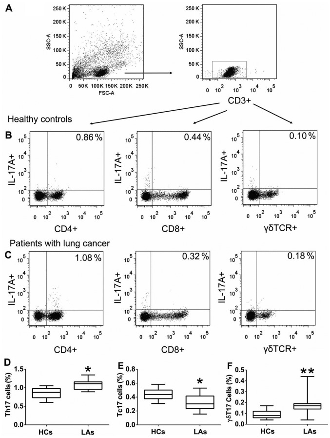
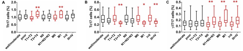
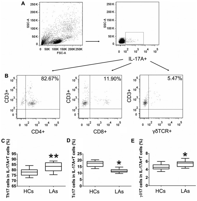
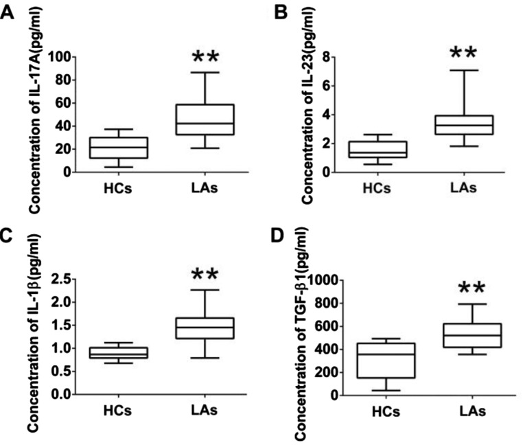
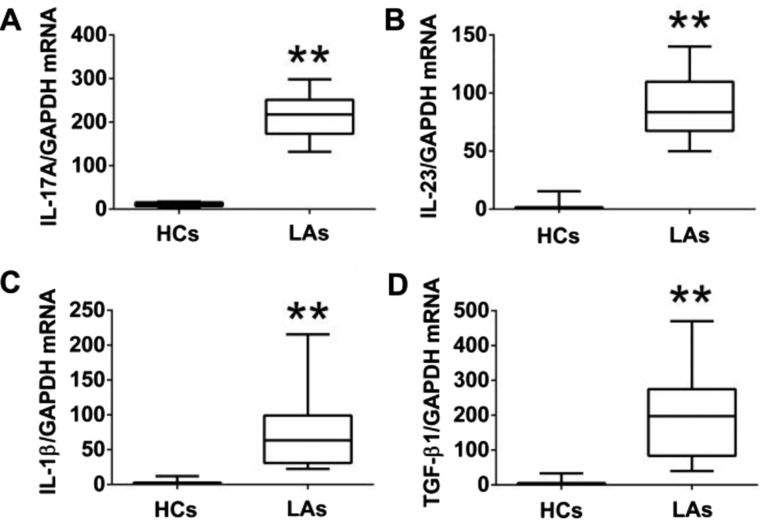
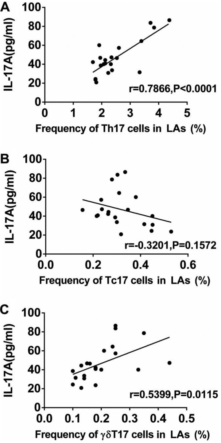
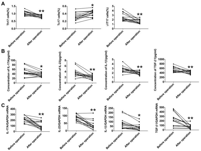
Accumulating evidence has shown that T cells are crucial in shaping the tumor microenvironment and regulating tumor development. However, the roles of IL-17A‑producing T cells (IL-17A+CD4+ Th17, IL-17A+CD8+ Tc17 and IL-17A+ γδT17 cells) and related cytokines in the progression of lung cancer (LC) remain uncertain. Here, we found that the frequencies of both Th17 and γδT17 cells in the peripheral blood of patients with lung adenocarcinoma (LA) were higher than those in healthy controls (HCs), whereas the frequency of Tc17 cells in the patients with LA was decreased. In addition, the frequencies of circulating Th17 and γδT17 cells, but not Tc17 cells, were positively associated with tumor invasion and metastasis. Furthermore, the major source of IL-17A production was Th17 cells, followed by Tc17 and γδT17 cells, in peripheral blood from patients with LA and HCs; but the percentages of Th17 and γδT17 cells in total intracellular IL-17A+ cells obtained from the patients with LC were higher than those from HCs. Moreover, the protein and corresponding mRNA levels of IL-17A, IL-23, IL-1β, and TGF-β1 were much higher in the patients with LA than those in HCs, and the levels of IL-17A in patients were positively correlated with numbers of both Th17 and γδT17 cells, but not Tc17 cells. Finally, the frequencies of circulating Th17 and γδT17 cells, along with the levels of IL-17A, IL-23, IL-1β, and TGF-β1 were decreased in the patients with LA after tumor resection, whereas the frequency of circulating Tc17 cells was inversely increased in these patients. Our findings indicate that Th17, Tc17, γδT17 cells, and IL-17A-associated cytokines contribute to the development of LA and thus represent promising targets for therapeutic strategies.


Figures

Figure 1
Figure 1
The frequency of circulating Th17, Tc17 and γδT17 cells in all individuals. Peripheral blood mononuclear cells (PBMCs) obtained from patients with lung adenocarcinoma (LA) and healthy controls (HCs) were stained with labeled antibodies and analyzed using flow cytometric analysis. (A) PBMCs were gated initially on living lymphocytes and then on CD3+ T cells. (B) Representative plots of IL-17A+CD4+ T (Th17), IL-17A+CD8+ T (Tc17) and, IL-17A+γδ+ T (γδT17) cells in total CD3+ T cells from HCs. (C) Representative plots of Th17, Tc17, and γδT17 cells in total CD3+ T cells from patients with LA. Comparison of (D) Th17, (E) Tc17, and (F) γδT17 cells presented as percentages of total CD3+ T cells, between patients with LA and HCs. The data shown represent the means ± SEM. *P<0.05, **P<0.01 were considered to represent significant differences compared with HCs.
Figure 2
Figure 2
Analysis of circulating Th17, Tc17 and γδT17 cells in patients with different tumor characteristics. The percentages of (A) Th17, (B) Tc17 and (C) γδT17 cells in patients with lung adenocarcinoma (LA) were compared according to tumor differentiation (good/moderate vs. poor), tumor invasion (T1+T2 vs. T3+T4), lymph node status (N0 vs. N1+N2+N3), metastasis (M0 vs. M1), and TNM stage (I+II and III+IV); The data shown represent the means ± SEM. *P<0.05, **P<0.01 were considered to represent significant differences.
Figure 3
Figure 3
Percentages of Th17, Tc17, and γδT17 cells in IL-17A+ cells. Peripheral blood mononuclear cells (PBMCs) obtained from patients with lung adenocarcinoma (LA) and healthy controls (HCs) were stained with labeled antibodies and analyzed using flow cytometric analysis. (A) PBMCs were gated initially on living lymphocytes and then on intracellular IL-17A. (B) Representative plots of Th17, Tc17, and γδT17 cells in IL-17A+ cells from patients with LA. Comparison of (C) Th17, (D) Tc17, and (E) γδT17 cells presented as percentages of total IL-17A+ cells between patients with LA and HCs. The data shown represent the means ± SEM. *P<0.05, **P<0.01 were considered to represent significant differences.
Figure 4
Figure 4
Protein levels of IL-17A and associated cytokines (IL-23, IL-1β and TGF-β1) in serum. Comparison of (A) IL-17A, (B) IL-23, (C) IL-1β, and (D) TGF-β1 concentration between patients with lung adenocarcinoma (LA) and healthy controls (HCs). The data shown represent the means ± SEM. *P<0.05, **P<0.01 were considered to represent significant differences.
Figure 5
Figure 5
The mRNA levels of IL-17A and associated cytokines (IL-23, IL-1β and TGF-β1) in PBMCs. Comparison of (A) IL-17A, (B) IL-23, (C) IL-1β, and (D) TGF-β1 mRNA levels between patients with lung adenocarcinoma (LA) and healthy controls (HCs). The data shown represent the means ± SEM. **P<0.01 was considered to represent significant differences.
Figure 6
Figure 6
Association between IL-17A protein levels and frequencies of circulating Th17, Tc17 and γδT17 cells in patients with lung adenocarcinoma (LA). (A) Association between the protein level of IL-17A and the frequencies of circulating (A) Th17, (B) Tc17, and (C) γδT17 cells. P<0.05 was considered to represent significant difference.
Figure 7
Figure 7
Alterations in IL-17A-producing T cells and IL-17A-associated cytokines in patients with lung adenocarcinoma (LA) after surgery. (A) Alterations in Th17, Tc17 and γδT17 cells in patients with LA after surgery. (B) Alterations in protein levels of IL-17A and associated cytokines (IL-23, IL-1β and TGF-β1) in patients with LA after thoracic surgery. (C) Alterations of mRNA levels of IL-17A, IL-23, IL-1β and TGF-β1 in patients with LA after surgery. *P<0.05, **P<0.01 were considered to represent significant differences.

References

    1. Islami F, Torre LA, Jemal A. Global trends of lung cancer mortality and smoking prevalence. Transl Lung Cancer Res. 2015;4:327–338. - PMC - PubMed
    1. Siegel RL, Miller KD, Jemal A. Cancer statistics, 2016. CA Cancer J Clin. 2016;66:7–30. doi: 10.3322/caac.21332. - DOI - PubMed
    1. She J, Yang P, Hong Q, Bai C. Lung cancer in China: Challenges and interventions. Chest. 2013;143:1117–1126. doi: 10.1378/chest.11-2948. - DOI - PubMed
    1. Hanahan D, Weinberg RA. Hallmarks of cancer: The next generation. Cell. 2011;144:646–674. doi: 10.1016/j.cell.2011.02.013. - DOI - PubMed
    1. Balkwill F, Mantovani A. Inflammation and cancer: Back to Virchow? Lancet. 2001;357:539–545. doi: 10.1016/S0140-6736(00)04046-0. - DOI - PubMed

MeSH terms