. 2016 Jun 9;11(1):43.
doi: 10.1186/s13024-016-0111-6.

Stathmin 1/2-triggered microtubule loss mediates Golgi fragmentation in mutant SOD1 motor neurons

Affiliations

Stathmin 1/2-triggered microtubule loss mediates Golgi fragmentation in mutant SOD1 motor neurons

Sarah Bellouze et al. Mol Neurodegener. .

Abstract

Background: Pathological Golgi fragmentation represents a constant pre-clinical feature of many neurodegenerative diseases including amyotrophic lateral sclerosis (ALS) but its molecular mechanisms remain hitherto unclear.

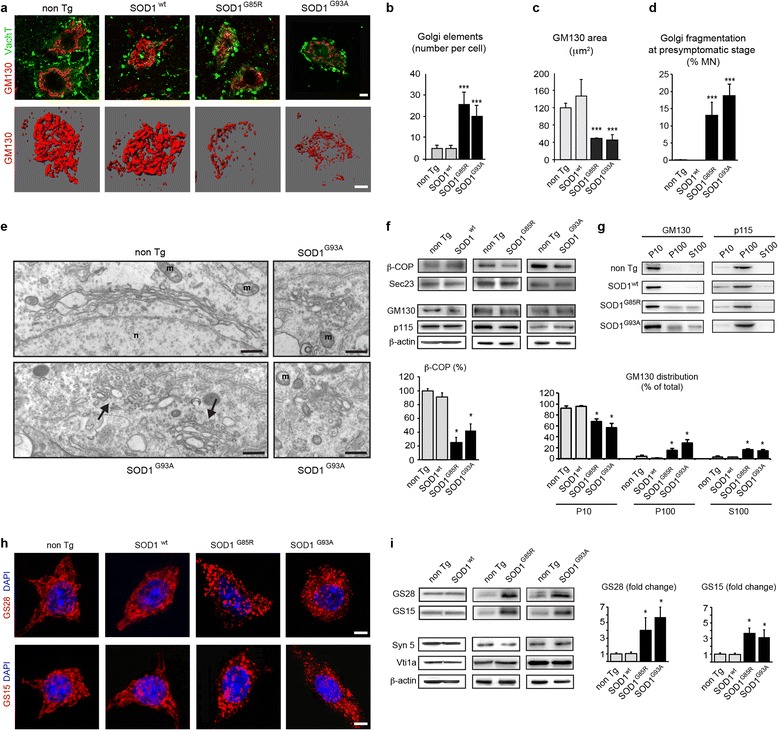
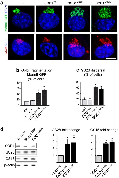
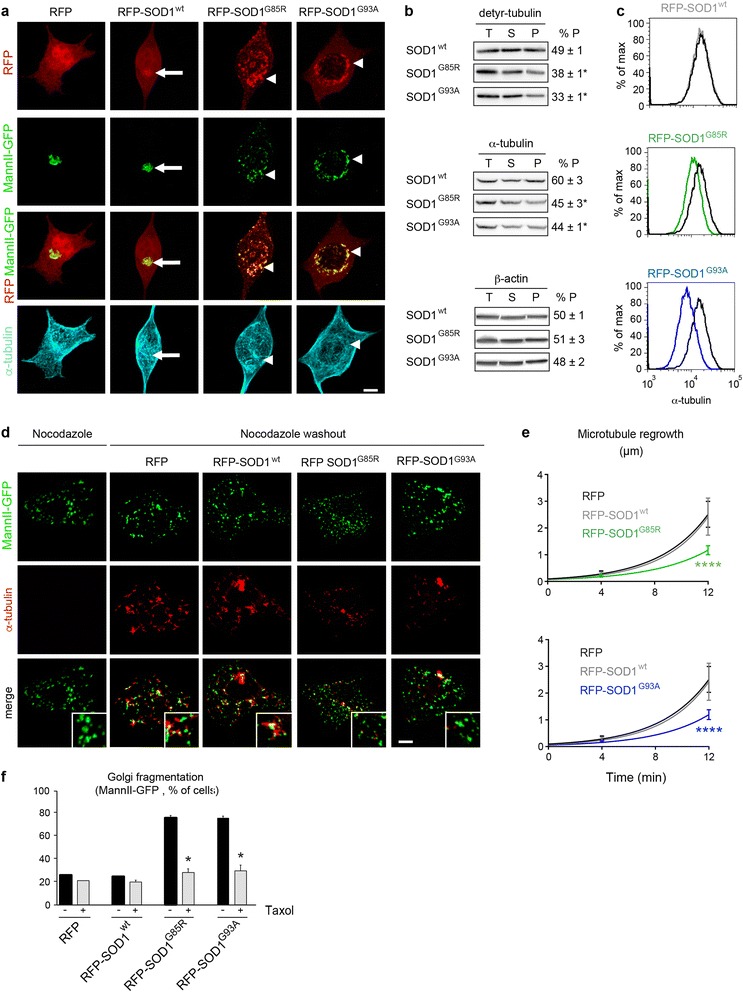
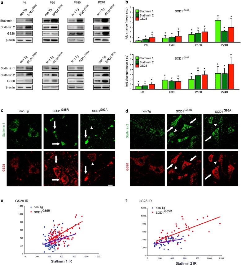
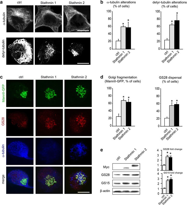
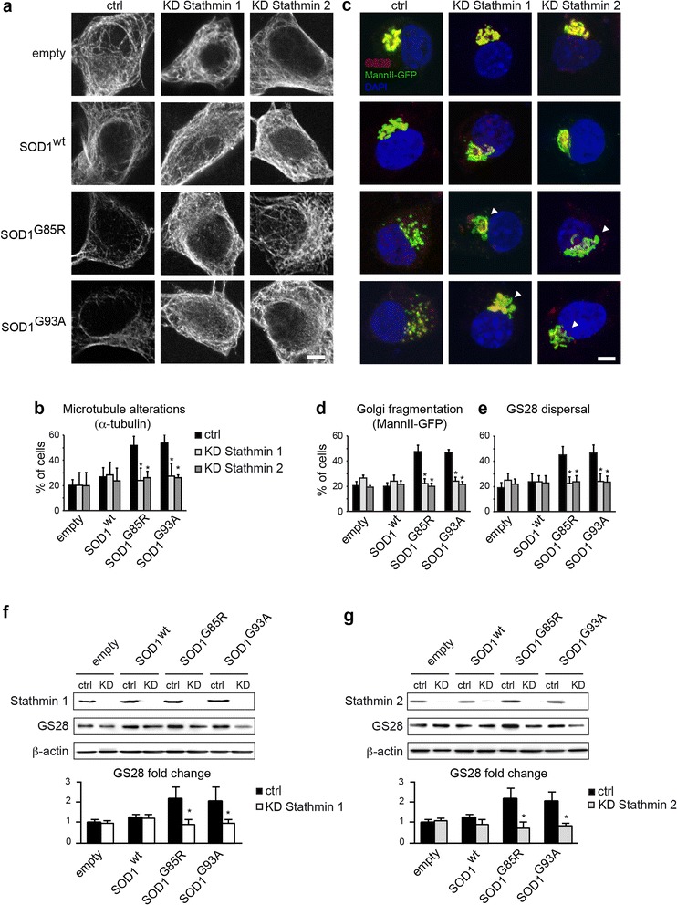
Results: Here, we show that the severe Golgi fragmentation in transgenic mutant SOD1(G85R) and SOD1(G93A) mouse motor neurons is associated with defective polymerization of Golgi-derived microtubules, loss of the COPI coat subunit β-COP, cytoplasmic dispersion of the Golgi tether GM130, strong accumulation of the ER-Golgi v-SNAREs GS15 and GS28 as well as tubular/vesicular Golgi fragmentation. Data mining, transcriptomic and protein analyses demonstrate that both SOD1 mutants cause early presymptomatic and rapidly progressive up-regulation of the microtubule-destabilizing proteins Stathmins 1 and 2. Remarkably, mutant SOD1-triggered Golgi fragmentation and Golgi SNARE accumulation are recapitulated by Stathmin 1/2 overexpression but completely rescued by Stathmin 1/2 knockdown or the microtubule-stabilizing drug Taxol.

Conclusions: We conclude that Stathmin-triggered microtubule destabilization mediates Golgi fragmentation in mutant SOD1-linked ALS and potentially also in related motor neuron diseases.


Figures

Fig. 1
Fig. 1
Morphological and molecular Golgi alterations in motor neurons of mutant SOD1 mice. a. Confocal z-stacks (upper panels) show fragmentation of GM130-labeled Golgi membranes in lumbar motor neurons of 240-day-old mutant SOD1G85R and SOD1G93A mice as compared to non-transgenic mice and transgenic SOD1wt mice. Motor neurons are identified by expression of VaChT (vesicular acetylcholine transporter). 3D-modeling (lower panels) of GM130-labelled Golgi membranes in entire motor neurons confirms Golgi fragmentation. Scale bars 10 μm. b. Increased number of GM130-stained Golgi elements in mutant SOD1G85R and SOD1G93A motor neurons determined by 3D modeling of Golgi membranes in entire cells (mean ± sd, n = 12 motor neurons from three mice per genotype, *** p < 0.0001 by student’s t-test, unpaired. c. Decreased cross-sectional area of GM130-labeled Golgi area in mutant SOD1G85R and SOD1G93A motor neurons as compared to control motor neurons (mean ± sd, *** p < 0.001, n = 50 motor neurons from three mice per genotype, student's t test). See also Additional file 1: Figure S1. d. Percentage of motor neurons with GM130-labeled Golgi fragmentation at presymptomatic stage (mean ± sd, *** p < 0.001 by student’s t-test, n > 250 motor neurons from four mice per genotype were analyzed at presymptomatic stage corresponding to age 130 days (SOD1G93A, non Tg) or 180 days (SOD1G85R, SOD1wt). e. Electron microscopy of a lumbar motor neuron from a non-transgenic mouse aged 140 days showing a typical Golgi apparatus (upper panel) that is easily distinguished from unlinked, partially swollen and vesiculated Golgi profiles observed in mutant SOD1G93A motor neurons (arrows in lower panel, magnifications on the right). n: nucleus, m: mitochondria. Scale bars 500 nm (left panels), 200 nm (right panels). f. Western blots showing decreased levels of β-COP in mutant SOD1 mice, and normal levels of Sec23, GM130 and p115. Loading control β-actin. Shown is one representative blot per genotype out of four. The diagram below shows that β-COP levels (normalized to β-actin) are reduced to 25 ± 7.7 % and 42.5 ± 9.6 % of non Tg (mean ± sd, n = 4 per genotype, * p < 0.01 by Mann Whitney test). g. Subcellular fractionation of spinal cords. Western blot analyses show redistribution of GM130 in mutant SOD1 mice, as indicated by shift from its normal membrane localization in control and SODwt mice into fragmented membranes and vesicles in SOD1G85R and SOD1G93A mice and cytosol. P115 is not redistributed. Each blot is representative of three independant experiments on mice of the indicated genotypes. The diagram below shows densitometric determination of protein distribution (mean ± sd, n = 3 per genotype, * p < 0.01 by Mann Whitney test). h. Confocal imaging reveals accumulation of Golgi v-SNARE protein GS28 (upper panels) and GS15 (lower panels) in small vesicle-like punctae of motor neurons in mutant SOD1 mice, as compared to controls. Scale bars 10 μm. i. Western blots (upper two panels) demonstrating massively increased levels of Golgi v-SNAREs GS28 and GS15 in mutant SOD1G85R and SOD1G93A lumbar spinal cords, as compared to non-transgenic and SOD1wt spinal cords. Western blots (lower three panels) showing normal expression of the Golgi t-SNARE Syntaxin-5a and the endosomal v-SNARE Vti1a. β-actin indicates equal protein loading. Each blot is representative of three independant experiments on mice of the indicated genotypes. The diagrams show increased spinal cord protein levels of GS28 by 4 ± 1.6 (SOD1G85R) and by 3.7 ± 1.3 (SOD1G93A) fold and of GS15 by 3.7 ± 0.7 (SOD1G85R) and by 3.2 ± 1 (SOD1G93A) as compared to non Tg control (mean ± sd, * p < 0.001 by Mann Whitney test)
Fig. 2
Fig. 2
Golgi alterations in mutant SOD1-transfected NSC-34 motor neurons. a. Confocal images (upper panels) showing Golgi fragmentation with the marker MannII-GFP in NSC-34 cells transfected for 4 DIV with mutant SOD1, as compared to the situation in control or wildtype SOD1-transfected cells. Confocal images (lower panels) showing GS28 dispersal after transfection with mutant SOD1, as compared to the situation in control or wildtype SOD1 cells. Scale bar 10 μm. b. Percentage of cells (mean ± sd) with Golgi fragmentation labeled for MannII-GFP. More than 300 cells per condition were analyzed on quadruplicate wells per condition. Statistical significance * p < 0.01 by Mann–Whitney test, as compared to mock and SODwt. Data represent one out of three experiments yielding similar results. c. Percentage of transfected cells with dispersal of GS28. More than 200 cells per condition were analyzed on quadruplicate wells per condition. Statistical significance * p < 0.01 by Mann–Whitney test, as compared to mock and SODwt. d. Western blot analysis of NSC-34 cells demonstrating that GS28 protein levels are increased to 2.7 ± 0.7 and 2.9 ± 0.8 by SOD1G85R and SOD1G93A, respectively, as compared to those in control SODwt. GS15 protein levels are increased to 2.7 ± 0.2 and 3.0 ± 0.3 fold, respectively. The diagrams show densitometric quantification of protein levels relative to SODwt (mean ± sd). Cellular extracts from three independently transfected cultures were blotted and analyzed each in duplicate. Statistical significance * p < 0.01 by Mann Whitney test
Fig. 3
Fig. 3
SOD1 mutants impair polymerization of Golgi-derived microtubules. a. Images show NSC-34 cells expressing RFP or SOD1 variants tagged with RFP after extraction for soluble proteins and labeling with MannII-GFP (Golgi) and antibodies against α-tubulin (microtubules). Merged RFP/MannII-GFP images show that both wildtype SOD1 (arrow) and mutant SOD1 (arrowheads) localize to Golgi membranes. This is confirmed by Pearson’s correlation analysis (RFP-SOD1wt 0.74 ± 0.09, RFP-SOD1G85R 0.72 ± 0.09, RFP-SOD1G93A 0.77 ± 0.14, RFP 0.13 ± 0.14, statistical significance SOD1 variants vs RFP : p < 0.0006 by student’s t-test). Mutant SOD1G85R and SOD1G93A specifically cause rarefaction of microtubules around fragmented Golgi profiles (arrowheads in lower panel). b. Biochemical fractionation of total (T), soluble (S) and polymerized (P) tubulins. Cells expressing SOD1G85R or SOD1G93A display a decreased ratio of polymerized detyrosinated (detyr-) tubulin. The ratio of polymerized α-tubulin is also decreased. Polymerization of β-actin is not affected by mutant SOD1. % P is equivalent to P/(P + S), * p < 0.01 by Mann–Whitney test, n = 3 experiments each. c. Flow cytometry of cellular microtubules in NSC-34 cells after extraction of soluble tubulins, microtubule stabilization and intracellular labeling with α-tubulin-FITC antibodies. Cells expressing SODwt (in grey, upper panel) display normal levels of α-tubulin-containing microtubules in comparison to cells expressing RFP (in black). Cells expressing mutant SOD1G85R (in green, middle panel) or mutant SOD1G93A (in blue, lower panel) show decreased levels of cellular microtubules. Median fluorescence signal per cell: 15.800 (RFP), 15.900 (SOD1wt), 11.600 (SOD1G85R), 8.418 (SOD1G93A). Statistical significance by chi square test, * T(x) > 200, ns T(x) = 0. d. Images showing transfected NSC-34 cells that were treated with Nocodazole (left column) and restored to drug-free medium for 12 min (right four columns). Cells were identified by RFP expression (not shown), Golgi profiles were identified by MannII-GFP and growing microtubules with antibodies against α-tubulin (pseudocolored in red). Mutant SOD1G85R and SOD1G93A impede regrowth of Golgi-derived microtubules (zoomed insets). Scale bar 5 μm. e. Diagrams showing reduced growth rate of Golgi-derived microtubules in cells expressing RFP- SOD1G85R (upper panel) or RFP- SOD1G93A (lower panel) as compared to cells expressing RFP- SOD1wt or RFP. Microtubule length represents mean of mean of >12 cells per time point and condition and a total of 1495 microtubules analyzed. Statistical significance **** p < 0.0001 (RFP-SOD1G85R vs RFP or vs SOD1wt) and **** p < 0.0001 (RFP-SOD1G93A vs RFP or vs SOD1wt) by ANOVA test and Tukey’s multiple comparison test. f. Diagrams showing Taxol-mediated rescue of MannII-GFP-labeled Golgi fragmentation in cells expressing mutant SOD1G85R or SOD1G93A each tagged to RFP. Statistical significance * p < 0.01 (Taxol vs mock) by Mann–Whitney test, n ≥ 50 cells per well and 4 replicate wells were analyzed per condition
Fig. 4
Fig. 4
Early and rapidly progressive co-accumulation of Stathmin 1, Stathmin 2 and GS28 in motor neurons of mutant SOD1 mice. a. Western blots show expression of Stathmin 1, Stathmin 2 and GS28 in lumbar spinal cords of SOD1G85R mice (upper panels), SOD1G93A mice (lower panels) and corresponding litter mates. Analyses were performed at ages P8, P30, P180 and P240. Loading control β-actin. Each blot was performed in duplicate and is representative of four animals per genotype. b. Diagrams showing kinetics of Stathmin 1, Stathmin 2 and GS28 protein levels in lumbar spinal cords of mutant SOD1G85R mice and SOD1G93A mice. Stathmin 1 and 2 levels are already significantly increased at P8. Fold changes (mean ± sd) are determined from four spinal cords per genotype and time point and expressed relative to the levels in non-transgenic littermate controls (set to 1). Differences between mutant SOD1 and control are statistically significant as measured by Mann Whitney test (*, p < 0.01). c-d. Confocal images of lumbar spinal cord cross sections from non-transgenic, SOD1G85R and SOD1G93A mice aged 240 days showing accumulation of Stathmin 1 (C, upper panels), Stathmin 2 (D, upper panels) and GS28 (C-D, lower panels) in motor neurons, which sometimes appear as degenerating. Note motor neurons with either low expression (arrowheads) or high expression (arrows) of Stathmins and Golgi SNAREs. Scale bar 20 μm. e. Diagram showing immunoreactivities (IR) of Stathmin 1 (x-axis) and GS28 (y-axis) in motor neurons of control non-transgenic mice (in blue) and mutant SOD1G85R mice (in red). Pearson analysis demonstrates significant correlation between both parameters: r = 0.28, p < 0.0066 (ctrl) and r = 0.47, p < 0.0001 (SOD1G85R). The slopes of the regression curves are 0.49 ± 0.18 (ctrl) and 0.68 ± 0.13 (SOD1G85R), n = 91 cells (ctrl) and n = 99 cells (SOD1G85R) analyzed from n = 3 mice per genotype. f. Immunoreactivities of Stathmin 2 and GS28 also show significant correlation by Pearson analysis: r = 0.40, p < 0.0047 (ctrl), r = 0.69, p < 0.0001 (SOD1G85R). The slopes of the regression curves are 0.29 ± 0.09 (ctrl) and 0.60 ± 0.09 (SOD1G85R) by linear regression analysis, n = 47 cells and n = 48 cells analyzed respectively from n = 3 mice per genotype
Fig. 5
Fig. 5
Stathmin 1/2 overexpression phenocopies expression of mutant SOD1. a. Images showing microtubule alterations in NSC-34 motor neurons transfected with Myc-tagged Stathmin 1 or Stathmin 2, as compared to control cells. Cells were cultured for 2 DIV, fixed with paraformaldehyde and analyzed with antibodies α-tubulin (upper panels), detyrosinated tubulin (lower panels) or Myc (not shown). Scale bar 5 μm. b. Quantitative analyses showing percentage of cells with rarefied or broken microtubules (α-tubulin or detyrosinated tubulin). Data represent one typical out of five independent experiments each done in quadruplicate per condition. Number of cells analyzed (ctrl, Stathmin 1, Stathmin 2): α-tubulin (175, 174, 139), detyr-tubulin (184, 146, 152). Statistical significance by Mann–Whitney test: * p < 0.001 as compared to mock. c. Images showing Golgi alterations (MannII-GFP, GS28) together with microtubule alterations (α-tubulin) in NSC-34 motor neurons transfected with Stathmin 1 or Stathmin 2 as compared to control cells. Scale bar 5 μm. d. Quantitative analyses showing percentage of cells with Golgi alterations (MannII-GFP) or GS28 dispersal). Data represent one typical out of five independent experiments each done in quadruplicate per condition. Number of cells analyzed (ctrl, Stathmin 1, Stathmin 2): MannII-GFP (>500 each). GS28 (482, 434, 526). Statistical significance by Mann–Whitney test: * p < 0.001 as compared to mock. e. Western blot analysis of cells transfected with plasmids encoding Myc-tagged Stathmin 1 or Stathmin 2 or with an empty control plasmid (pcDNA). GS15 and GS28 levels are increased after overexpression of Stathmin 1 by 2.7 ± 0.3 and 2.6 ± 0.3 (mean ± sd) respectively. A similar increase in Golgi SNAREs is observed after overexpression of Stathmin 2 (GS15 2.6 ± 0.2, GS28 2.9 ± 0.3). n = 4 blots corresponding to independent experiments, * p < 0.01 by Mann - Whitney test
Fig. 6
Fig. 6
Stathmin 1/2 knockdown rescues microtubule and Golgi alterations triggered by mutant SOD1. a. Confocal images showing NSC-34 cells transfected with empty, SOD1wt, SOD1G85R or SOD1G93A plasmids as well as ctrl, Stathmin-1 or Stathmin-2 siRNAs. Cells were immunolabelled for α-tubulin. Note that knockdown of Stathmin 1 or 2 rescues mutant SOD1-triggered microtubule alterations. Transfected cells were identified by expression of co-transfected MannII-GFP (not shown). Scale bar 5 μm. b. Diagram showing percentage of microtubule alterations (mean ± sd) under the different conditions. >150 cells were analyzed per condition. Statistical significance * p < 0.01 by Mann–Whitney test. Data are representative of three independent experiments. c. Confocal images showing NSC-34 cells transfected as in A and counterstained for GS28. Note rescue (arrowheads) of mutant SOD1-triggered Golgi fragmentation and GS28 dispersal by knockdown of Stathmin 1 or 2. Scale bar 5 μm. d-e. Diagram showing percentage (mean ± sd) of Golgi alterations labelled by MannII-GFP (d) or GS28 (e) under the different conditions. >150 cells were analyzed per condition. Statistical significance * p < 0.01 by Mann–Whitney test. Data are representative of four independent experiments. f-g. Western blots show efficient siRNA-mediated knockdown (KD) of Stathmin 1 (f) and Stathmin 2 (g) in NSC-34 cells transfected with empty, SOD1wt, SOD1G85R or SOD1G93A plasmids as compared to ctrl siRNA (upper blots). Both Stathmin-1 or Stathmin-2 knockdown reduces the GS28 up-regulation triggered by SOD1G85R or SOD1G93A (middle blots). Loading control β-actin (lower blots) indicates reduced protein content in lanes 1 and 2 in F (empty ctrl, empty Stathmin-1 KD). The diagrams below show densitometric quantification of GS28 protein levels each normalized to β-actin (n = 4 blots, mean ± sd, * statistically significant differences between ctrl siRNA and Stathmin 1/2 siRNA as measured by Mann - Whitney test)

References

    1. Acevedo-Arozena A, Kalmar B, Essa S, Ricketts T, Joyce P, Kent R, Rowe C, Parker A, Gray A, Hafezparast M, et al. A comprehensive assessment of the SOD1G93A low-copy transgenic mouse, which models human amyotrophic lateral sclerosis. Dis model Mech. 2011;4:686–700. doi: 10.1242/dmm.007237. - DOI - PMC - PubMed
    1. Aguilera-Gomez A, Rabouille C. Intra Golgi transport. In: Bradshaw R, Stahl P (eds) Encyclopedia of Cell Biology. Elsevier; 2015; 354-362. doi:10.1016/B978-0-12-394447-4.20034-5.
    1. Amayed P, Pantaloni D, Carlier MF. The effect of stathmin phosphorylation on microtubule assembly depends on tubulin critical concentration. J Biol Chem. 2002;277:22718–22724. doi: 10.1074/jbc.M111605200. - DOI - PubMed
    1. Atkin JD, Farg MA, Soo KY, Walker AK, Halloran M, Turner BJ, Nagley P, Horne MK. Mutant SOD1 inhibits ER-Golgi transport in amyotrophic lateral sclerosis. J Neurochem. 2014;129:190–204. doi: 10.1111/jnc.12493. - DOI - PubMed
    1. Bellouze S, Schaefer MK, Buttigieg D, Baillat G, Rabouille C, Haase G. Golgi fragmentation in pmn mice is due to a defective ARF1/TBCE cross-talk that coordinates COPI vesicle formation and tubulin polymerization. Hum Mol Genet. 2014;23:5961–5975. doi: 10.1093/hmg/ddu320. - DOI - PubMed

Publication types

LinkOut - more resources