. 2016 Jun 8;36(23):6312-20.
doi: 10.1523/JNEUROSCI.0245-16.2016.

Perineuronal Nets Suppress Plasticity of Excitatory Synapses on CA2 Pyramidal Neurons

Affiliations

Perineuronal Nets Suppress Plasticity of Excitatory Synapses on CA2 Pyramidal Neurons

Kelly E Carstens et al. J Neurosci. .

Abstract

Long-term potentiation of excitatory synapses on pyramidal neurons in the stratum radiatum rarely occurs in hippocampal area CA2. Here, we present evidence that perineuronal nets (PNNs), a specialized extracellular matrix typically localized around inhibitory neurons, also surround mouse CA2 pyramidal neurons and envelop their excitatory synapses. CA2 pyramidal neurons express mRNA transcripts for the major PNN component aggrecan, identifying these neurons as a novel source for PNNs in the hippocampus. We also found that disruption of PNNs allows synaptic potentiation of normally plasticity-resistant excitatory CA2 synapses; thus, PNNs play a role in restricting synaptic plasticity in area CA2. Finally, we found that postnatal development of PNNs on CA2 pyramidal neurons is modified by early-life enrichment, suggesting that the development of circuits containing CA2 excitatory synapses are sensitive to manipulations of the rearing environment.

Significance statement: Perineuronal nets (PNNs) are thought to play a major role in restricting synaptic plasticity during postnatal development, and are altered in several models of neurodevelopmental disorders, such as schizophrenia and Rett syndrome. Although PNNs have been predominantly studied in association with inhibitory neurons throughout the brain, we describe a dense expression of PNNs around excitatory pyramidal neurons in hippocampal area CA2. We also provide insight into a previously unrecognized role for PNNs in restricting plasticity at excitatory synapses and raise the possibility of an early critical period of hippocampal plasticity that may ultimately reveal a key mechanism underlying learning and memory impairments of PNN-associated neurodevelopmental disorders.

Keywords: critical period; extracellular matrix; hippocampus; long-term potentiation.


Figures

Figure 1.
Figure 1.
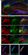
PNN markers surround CA2 pyramidal neurons and dendrites. a, Fluorescent labeling of WFA (green) localizes with CA2 pyramidal neurons and with GAD67-positive inhibitory neurons (red) in hippocampus. b, Anti-aggrecan (top), WFA (middle), and neurocan (bottom), indicated in green, label EGFP-expressing CA2 pyramidal neurons and their proximal neurites in red (scale bar, 50 μm). c, Fluorescent ISHs showing that aggrecan mRNA (green) and a CA2 marker, Pcp4 mRNA (red), colocalize to the CA2 pyramidal cell layer. Yellow shows the overlapping distribution of these two mRNAs.
Figure 2.
Figure 2.
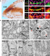
PNN markers are associated with excitatory synapses in area CA2. a, WFA immunoperoxidase staining in area CA2 labels CA2 dendrites in the SR in addition to cell bodies in the SP. b, WFA label surrounds excitatory synapses on primary apical dendrites of area CA2 SR; green is GFP expressed in neurons in tissue from a thy1-GFP-M mouse, red is WFA, purple is VGLUT1, and yellow shows where two channels depicted in each panel have overlapped (scale bar, 1 μm). c, Electron micrographs showing WFA staining along dendritic spines of CA2 pyramidal neurons in the SR (top, red arrowheads), and area CA2 somata in the SP (bottom), and sparse labeling of WFA in area CA1 SP and SR (bottom; scale bar, 2 μm).
Figure 3.
Figure 3.
PNNs increase during postnatal development and are increased with experience. a, WFA staining in area CA2 is increased in animals reared in an enriched environment, compared with animals raised in standard (control) cages. Note that the images have been digitally lightened in all panels equally to better display PNNs at PN 14 in area CA2. b, Normalized WFA fluorescence intensity was significantly greater in CA2 SP of EE mice compared with control at PN 21 (N = 8 and 7 for control and EE respectively) and PN 45 (N = 6 and 9 for control and EE respectively; Bonferroni's post hoc test for pairwise comparison after two-way ANOVA,*p < 0.05, ***p = 0.0008). No significant difference was observed at PN 14 (N = 4 and 5 for control and EE respectively). A two-way ANOVA for the two conditions at different ages indicated significant main effects of age (F(2,33) = 32.33), condition (F(2,33) = 11.72), and interaction (F(2,33) = 3.338). Indicated is the mean ± SEM. c, Normalized WFA fluorescence intensity was similarly significantly greater in area CA2 SR at PN 21 and 45 (same N's as reported in b; Bonferroni post hoc test for pairwise comparison after two-way ANOVA,*p < 0.05, ***p = 0.0009). A two-way ANOVA for the two conditions at different ages indicated significant main effects of age (F(2,33) = 69.55), condition (F(1,33) = 11.93), and interaction (F(2,33) = 3.940).
Figure 4.
Figure 4.
Disruption of PNNs allows for potentiation of EPSCs in CA2 neurons. a, PNNs are degraded in 300-μm-thick hippocampal slices cleared with thiodiethanol after a 2 h incubation with ChABC compared with control; fluorescent labeling of PNNs with WFA (green) and CA2 neurons with the Amigo2-EGFP mouse (red). Slices shown are from animals at PN 14. b, Plasticity of EPSC amplitudes is enhanced in CA2 neurons treated with the PNN-degrading enzyme ChABC, compared with untreated controls. After baseline, an LTP pairing protocol (270 pulses at 3 Hz paired with postsynaptic depolarization; at time 0), resulted in potentiation of EPSCs in CA2 neurons treated with 0.05 U/ml ChABC (mean over 22–28 min, n = 10), but not in untreated CA2 controls (n = 10). One-way ANOVA, Bonferroni's post hoc test for pairwise comparison, *p < 0.05. Indicated is the mean ± SEM. LTP induced in CA1 neurons is shown for comparison. Insets are representative traces of EPSCs from CA2 control and CA2 ChABC-treated neurons before and 20 min after the LTP pairing protocol. c, ChABC treatment did not significantly alter action potential firing frequency in response to indicated current injection (n = 15 per group; two-way ANOVA, p > 0.05). Example traces show action potential firing of control (untreated) CA2 neuron (40 pA current steps from −100 to 180 pA displayed). d, e, ChABC did not significantly alter excitatory current amplitude (n = 6 per group) or paired-pulse ratio in CA2 neurons (S1, peak of first stimulus response; S2, peak of second stimulus response; n = 10 ChABC-treated neurons and n = 5 control; two-way ANOVA, Bonferroni's post hoc test). f, ChABC did not significantly alter AMPAR/NMDAR ratio measured at +40 mV (left) or at −70 mV (n = 6 ChABC-treated neurons and n = 8 controls; two-tailed unpaired t test). Measurements in d–f were made in the presence of a GABAA antagonist bicuculline; see Materials and Methods for details. Insets are representative traces of EPSCs from CA2 control holding at −70 and +40 mV.

References

    1. Alpár A, Gärtner U, Härtig W, Brückner G. Distribution of pyramidal cells associated with perineuronal nets in the neocortex of rat. Brain Res. 2006;1120:13–22. doi: 10.1016/j.brainres.2006.08.069. - DOI - PubMed
    1. Aoyagi Y, Kawakami R, Osanai H, Hibi T, Nemoto T. A rapid optical clearing protocol using 2,2′-thiodiethanol for microscopic observation of fixed mouse brain. PloS One. 2015;10:e0116280. doi: 10.1371/journal.pone.0116280. - DOI - PMC - PubMed
    1. Baroncelli L, Scali M, Sansevero G, Olimpico F, Manno I, Costa M, Sale A. Experience affects critical period plasticity in the visual cortex through an epigenetic regulation of histone post-translational modifications. J Neurosci. 2016;36:3430–3440. doi: 10.1523/JNEUROSCI.1787-15.2016. - DOI - PMC - PubMed
    1. Bartoletti A, Medini P, Berardi N, Maffei L. Environmental enrichment prevents effects of dark-rearing in the rat visual cortex. Nat Neurosci. 2004;7:215–216. doi: 10.1038/nn1201. - DOI - PubMed
    1. Belichenko PV, Hagberg B, Dahlström A. Morphological study of neocortical areas in Rett syndrome. Acta Neuropathol. 1997;93:50–61. doi: 10.1007/s004010050582. - DOI - PubMed

Publication types

MeSH terms