. 2016 Jun 9:6:27540.
doi: 10.1038/srep27540.

Pharmacological exploitation of the phenothiazine antipsychotics to develop novel antitumor agents-A drug repurposing strategy

Affiliations

Pharmacological exploitation of the phenothiazine antipsychotics to develop novel antitumor agents-A drug repurposing strategy

Chia-Hsien Wu et al. Sci Rep. .

Abstract

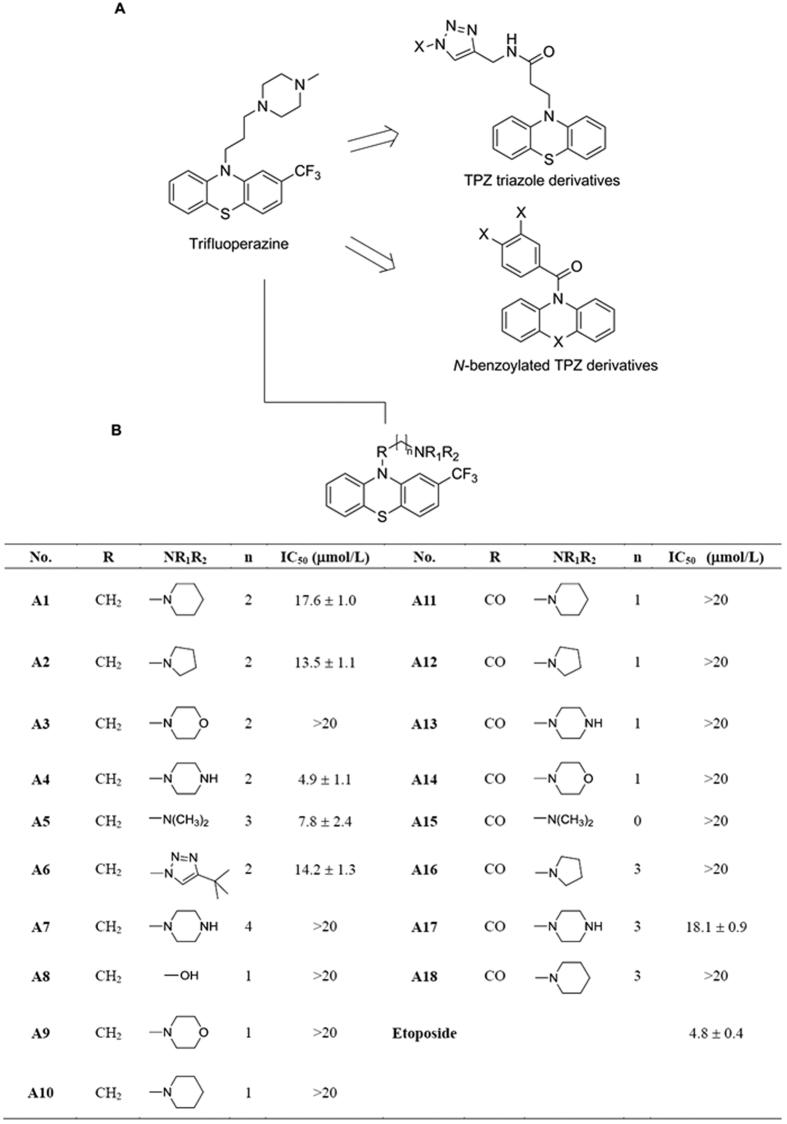
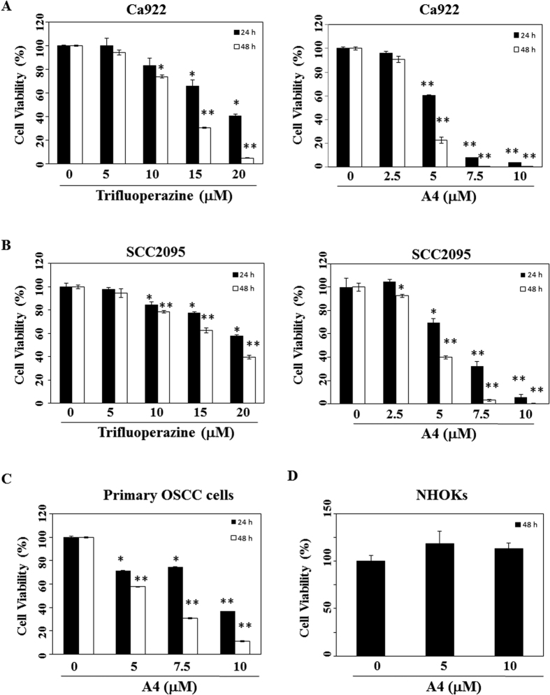
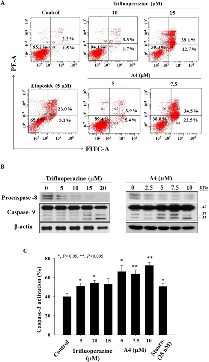
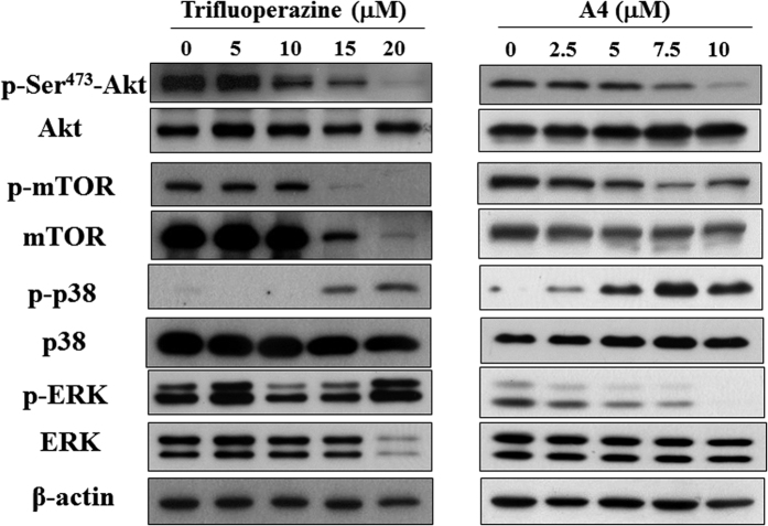
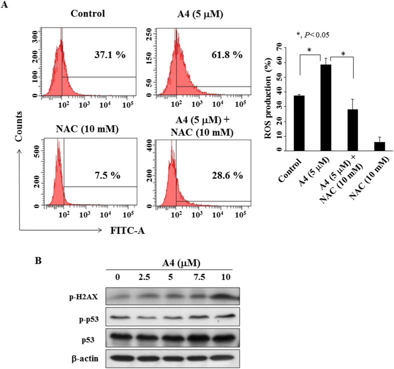
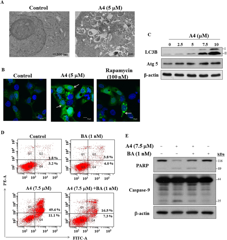
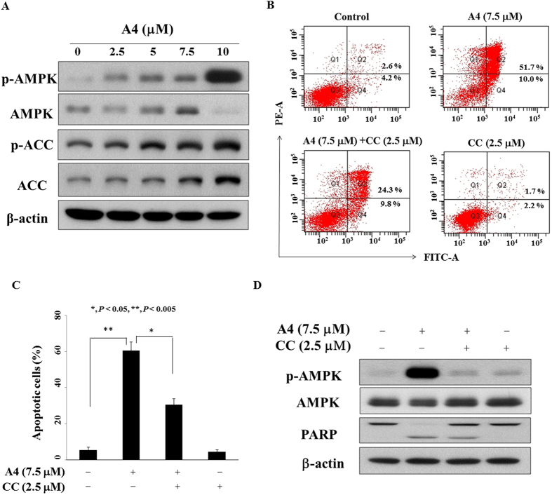
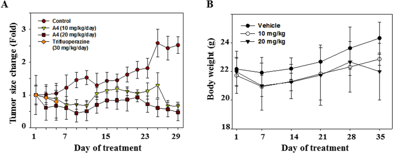
Phenothiazines (PTZs) have been used for the antipsychotic drugs for centuries. However, some of these PTZs have been reported to exhibit antitumor effects by targeting various signaling pathways in vitro and in vivo. Thus, this study was aimed at exploiting trifluoperazine, one of PTZs, to develop potent antitumor agents. This effort culminated in A4 [10-(3-(piperazin-1-yl)propyl)-2-(trifluoromethyl)-10H-phenothiazine] which exhibited multi-fold higher apoptosis-inducing activity than the parent compound in oral cancer cells. Compared to trifluoperazine, A4 demonstrated similar regulation on the phosphorylation or expression of multiple molecular targets including Akt, p38, and ERK. In addition, A4 induced autophagy, as evidenced by increased expression of the autophagy biomarkers LC3B-II and Atg5, and autophagosomes formation. The antitumor activity of A4 also related to production of reactive oxygen species and adenosine monophosphate-activated protein kinase. Importantly, the antitumor utility of A4 was extended in vivo as it, administrated at 10 and 20 mg/kg intraperitoneally, suppressed the growth of Ca922 xenograft tumors. In conclusion, the ability of A4 to target diverse aspects of cancer cell growth suggests its value in oral cancer therapy.


Figures

Figure 1
Figure 1. Use of trifluoperazine as scaffolds for developing new anticancer agents.
Upper panel, Structure of trifluoperazine, Lower panel, structures and potencies for inducing apoptotic death in Ca922 cells of the trifluoperazine derivatives A1 to A18. Cell viability was assessed by MTT assays with six replicates. The reported IC50 values are concentrations at which Ca922 cell death measures 50% relative to DMSO control after 48 h exposure in 5% FBS-containing MEM in 96-well plates.
Figure 2
Figure 2. Antiproliferative effects of trifluoperazine and A4 in oral cancer cell lines (Ca922, SCC2095), primary OSCC cells, and NHOKs.
(A) Ca922, (B) SCC2095 (C) Primary OSCC cells, and (D) NHOKs (5 × 103/200 μL) were treated with DMSO vehicle or trifluoperazine or A4 at the indicated concentrations. Cell viability was assessed by MTT assay as described in the Material section. Points, mean; bars, S.D. (n = 6). *P < 0.05, **P < 0.005 compared to the control group.
Figure 3
Figure 3. Evidence of apoptosis for trifluoperazine and A4-induced cell death.
(A) Annexin V-FITC/propidium iodide staining. Ca922 cells were treated with DMSO vehicle or trifluoperazine or A4 at the indicated concentrations in 5% FBS-supplemented MEM medium for 48 h. (B) Western blotting of procaspase-8 and caspase-9 after the treatment of trifluoperazine or A4 in Ca922 cells for 48 h. (C) Caspase-3 activation of trifluoperazine and A4 in Ca922 cells (n = 4). Staurosporine (Stauro.) as the positive control. *P < 0.05; **P < 0.005 compared to the control group.
Figure 4
Figure 4. Dose-dependent effects of trifluoperazine and A4 on the phosphorylation/expression of Akt, mTOR, p38, and ERK in Ca922 cells.
Cells are treated with trifluoperazine or A4 at the indicated concentrations in 5% FBS-MEM for 48 h and cell lysates were immunoblotted as described in Methods section.
Figure 5
Figure 5. ROS generation of A4 in Ca922 cells.
(A) Left panel, Flow cytometric analysis of the effect of A4 (5 μM), alone or in combination with the antioxidant N-acetylcysteine (NAC) for 3 h on ROS production. Three independent experiments were performed, and the statistical analysis are presented in right panel, Points, mean; bars, S.D. (n = 3). *P < 0.05 compared to the control group. (B) Western blotting analysis of the phosphorylation/expression of H2AX and p53 in Ca922 cells.
Figure 6
Figure 6. A4 induced autophagy.
(A) Electron microscopic analysis of autophagosome formation after the treatment of A4 (5 μM) or DMSO in Ca922 cells for 24 h as described in Methods section. Magnification, 12000x. Arrow: autophagosomes. (B) Fluorescent confocal microscopic analysis of A4-induced autophagosome formation in Ca922 cells ectopically expressing GFP-LC3. Cells transiently transfected with GFP-LC3 plasmids were treated with DMSO, 5 μM A4, or 100 nM rapamycin for 48 h and then fixed by 3.7% paraldehyde and examined by confocal microscopy. Scale bar: 10 μm. Arrow: autophagosomes. (C) Western blotting of LC3B and Atg5 in Ca922 cells treated with A4 for 48 h. (D) Ca922 cells were treated with 7.5 μM A4 alone or in combination with 1 nM bafilomycin A1 (BA) for 48 h, and then annexin V-FITC/PI double-staining analysis was performed. (E) Western blot analysis of the expression of PARP and caspase-9 after A4 alone or the combination of BA in Ca922 cells.
Figure 7
Figure 7. Restoration of antiproliferative activity of A4 by inactivating AMPK.
(A) The phosphorylation/expression of AMPK and ACC of A4 in Ca922 cells. Cells were treated with A4 in 5% FBS-supplemented MEM medium for 48 h, and cell lysates were immunoblotted as described in Methods. (B) Histogram showing 7.5 μM A4 alone or in combination with 2.5 μM compound c (CC) for 48 h, and then annexin V-FITC/PI double-staining analysis was performed. (C) The percentage of cells in Q2 and Q4 phases after the treatment was shown. Data are presented as mean ± S.D. *P < 0.05; **P < 0.005. (D) Western blotting analysis of the phosphorylation/expression of AMPK and PARP after the combination of CC or A4 alone in Ca922 cells.
Figure 8
Figure 8. Effects of A4 on Ca922 xenograft tumor size and mice body weight change.
(A) Mice bearing Ca922 xenografts were treated with normal saline control, A4 (10 mg/kg/day), A4 (20 mg/kg/day), or trifluoperazine (30 mg/kg/day). The tumor size was recorded every 2 days. (B) Body weight change of mice.

References

    1. Argiris A., Karamouzis M. V., Raben D. & Ferris R. L. Head and neck cancer. Lancet 371, 1695–1709 (2008). - PMC - PubMed
    1. Belcher R., Hayes K., Fedewa S. & Chen A. Y. Current treatment of head and neck squamous cell cancer. J Surg Oncol 110, 551–574 (2014). - PubMed
    1. Ohlow M. J. & Moosmann B. Phenothiazine: the seven lives of pharmacology’s first lead structure. Drug Discov Today 16, 119–131 (2011). - PubMed
    1. Jaszczyszyn A. et al. Chemical structure of phenothiazines and their biological activity. Pharmacol Rep 64, 16–23 (2012). - PubMed
    1. Sudeshna G. & Parimal K. Multiple non-psychiatric effects of phenothiazines: a review. Eur J Pharmacol 648, 6–14 (2010). - PubMed

Publication types

MeSH terms