Review
. 2016 Sep;173(18):2671-701.
doi: 10.1111/bph.13533. Epub 2016 Aug 10.

The function and regulation of acid-sensing ion channels (ASICs) and the epithelial Na(+) channel (ENaC): IUPHAR Review 19

Affiliations
Review

The function and regulation of acid-sensing ion channels (ASICs) and the epithelial Na(+) channel (ENaC): IUPHAR Review 19

Emilie Boscardin et al. Br J Pharmacol. 2016 Sep.

Abstract

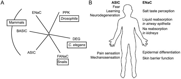
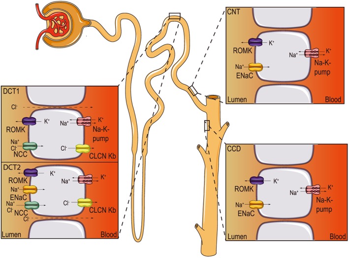
Acid-sensing ion channels (ASICs) and the epithelial Na(+) channel (ENaC) are both members of the ENaC/degenerin family of amiloride-sensitive Na(+) channels. ASICs act as proton sensors in the nervous system where they contribute, besides other roles, to fear behaviour, learning and pain sensation. ENaC mediates Na(+) reabsorption across epithelia of the distal kidney and colon and of the airways. ENaC is a clinically used drug target in the context of hypertension and cystic fibrosis, while ASIC is an interesting potential target. Following a brief introduction, here we will review selected aspects of ASIC and ENaC function. We discuss the origin and nature of pH changes in the brain and the involvement of ASICs in synaptic signalling. We expose how in the peripheral nervous system, ASICs cover together with other ion channels a wide pH range as proton sensors. We introduce the mechanisms of aldosterone-dependent ENaC regulation and the evidence for an aldosterone-independent control of ENaC activity, such as regulation by dietary K(+) . We then provide an overview of the regulation of ENaC by proteases, a topic of increasing interest over the past few years. In spite of the profound differences in the physiological and pathological roles of ASICs and ENaC, these channels share many basic functional and structural properties. It is likely that further research will identify physiological contexts in which ASICs and ENaC have similar or overlapping roles.


Figures

Figure 1
Figure 1
Relations and roles of ENaC and ASICs. (A) Phylogenetic tree of the ENaC/degenerin (DEG) family, showing besides ASIC and ENaC the subfamilies pickpocket (PPK), degenerin, the FMRFa‐activated channel FaNaC and the BASIC (also known as hINaC or BLINaC). (B) Illustration of the different physiological and pathological roles of ASICs and ENaC.
Figure 2
Figure 2
ASIC and ENaC subunit organization. (A) Schematic view of one subunit in the context of the trimeric ASIC, highlighting the different domains finger (purple), knuckle (turquoise), β‐ball (orange), palm (yellow), thumb (blue) and transmembrane domains (red). (B) Structure of an ASIC1 subunit based on the crystal structure obtained from chicken ASIC1 binding Mit‐Tx (Baconguis et al., 2014). The domains are coloured as in (A). (C) Model of the extracellular part of αENaC (Kashlan and Kleyman, 2011). Colouring as in (A); the transmembrane part was not modeled.
Figure 3
Figure 3
Functional properties of ASIC. (A) Action potential induction by extracellular acidification to pH 6.4, mediated by ASICs, measured by whole‐cell current‐clamp from a mouse hippocampal neuron. (B) A pH 5‐induced current recorded in whole‐cell voltage clamp to −60 mV from a Chinese hamster ovary cell stably transfected with ASIC1a. (C) The pH‐dependence of steady‐state desensitization and of activation of ASIC1a. In steady‐state desensitization experiments, cells were exposed for 55 s to the indicated conditioning pH, and a stimulation pH 6 solution was applied for 5 s to open the not yet desensitized channels. The normalized current response at pH 6 is plotted as a function of the conditioning pH. For ASIC1a activation, the cells were perfused by a pH 7.4 solution, and once per minute, this solution was changed to one of acidic pH to open the channels. The normalized current is plotted as a function of the stimulation pH.
Figure 4
Figure 4
Extracellular pH changes during neurotransmission in the CNS. Illustration based on experiments performed in hippocampal, photoreceptor, amygdala and calyx synapses, showing a synapse between a pre‐ and postsynaptic neuronal terminal and an astrocyte. For simplification, only the transport of protons and of HCO3 is shown in the figure (grey arrows; protons are red dots). The black arrows indicate the activation of synaptic vesicle release by calcium entry. The numbers 1–3 (coloured in blue) represent potential mechanisms for the initial alkaline shift recorded in hippocampal synapses: neurotransmission stimulates calcium signalling in the perisynaptic cells. As a consequence, the mechanisms of calcium buffering system are stimulated. One of these mechanisms involves the PMCA, which pumps the accumulated intracellular calcium ions out of the cell in exchange for external protons, leading to extracellular alkalinization. The numbers 4–6 (coloured in red) represent potential mechanisms underlying the acidic shift. The glial NBC causes a gradual extracellular acidification (4). Intense neurotransmission increases astrocytic energy demand, resulting in lactate and CO2 production (5). For clarity, only the lactate shuttle into presynaptic terminals is shown. Lactate is transported outside the astrocyte by the H+‐coupled monocarboxylate transporter (MCT) and CO2 can freely diffuse, also leading to extracellular acidification at the synaptic cleft. Fast acidifications at the synaptic cleft may also occur as a result of synaptic vesicle release and also by non‐vesicular release involving an undefined transporter (6).
Figure 5
Figure 5
Extracellular pH‐sensitive ion channels in the PNS. Illustration based on experiments performed in small‐diameter DRG neurons innervating peripheral organs. The scheme indicates the pathway of a signal from the sensory nerve terminals in an organ (in the example the heart) to the spinal cord and the brain and focuses on two specific locations, the peripheral nerve terminal (left) and the signalling along the sensory nerve axon (right). Only the ion channels that are modulated by pHe on the nerve terminal and along the axon are shown, indicated by the symbols ‘+’ (stimulation by lowering of pHe) and ‘−‘ (inhibition by lowering of pHe). Red symbols indicate that this modulation increases excitability, and blue symbols indicate that it decreases excitability. The number of symbols correlates with the pH‐dependence of the regulation (Table 3) as indicated at the bottom of the figure.
Figure 6
Figure 6
ENaC expression along the aldosterone‐sensitive distal nephron (ASDN). Schematic view of a nephron, with detailed views of the ion transport mechanisms in cells of the DCT, CNT and CCD. CLCN Kb, voltage‐sensitive chloride channel Kb; ROMK, renal outer medullary potassium channel.

Similar articles

Cited by

References

    1. Abdrakhmanova G, Cleemann L, Lindstrom J, Morad M (2004). Differential modulation of beta2 and beta4 subunits of human neuronal nicotinic acetylcholine receptors by acidification. Mol Pharmacol 66: 347–355. - PubMed
    1. Abdrakhmanova G, Dorfman J, Xiao Y, Morad M (2002). Protons enhance the gating kinetics of the alpha3/beta4 neuronal nicotinic acetylcholine receptor by increasing its apparent affinity to agonists. Mol Pharmacol 61: 369–378. - PubMed
    1. Adams CM, Anderson MG, Motto DG, Price MP, Johnson WA, Welsh MJ (1998). Ripped pocket and pickpocket, novel drosophila deg/enac subunits expressed in early development and in mechanosensory neurons. J Cell Biol 140: 143–152. - PMC - PubMed
    1. Adams CM, Snyder PM, Welsh MJ (1999). Paradoxical stimulation of a DEG/ENaC channel by amiloride. J Biol Chem 274: 15500–15504. - PubMed
    1. Alexander SPH, Catterall WA, Kelly E, Marrion N, Peters JA, Benson HE et al. (2015a). THE CONCISE GUIDE TO PHARMACOLOGY 2015/16: voltage‐gated ion channels. Brit J Pharmacol 172: 5904–5941. - PMC - PubMed

Publication types

Substances

LinkOut - more resources