. 2016 Jun 8;13(1):28.
doi: 10.1186/s12989-016-0140-x.

Gender differences in murine pulmonary responses elicited by cellulose nanocrystals

Affiliations

Gender differences in murine pulmonary responses elicited by cellulose nanocrystals

Anna A Shvedova et al. Part Fibre Toxicol. .

Abstract

Background: Cellulose-based materials have been used for centuries to manufacture different goods derived from forestry and agricultural sources. In the growing field of nanocellulose applications, its uniquely engineered properties are instrumental for inventive products coming to competitive markets. Due to their high aspect ratio and stiffness, it is speculated that cellulose nanocrystals (CNC) may cause similar pulmonary toxicity as carbon nanotubes and asbestos, thus posing a potential negative impact on public health and the environment.

Methods: The present study was undertaken to investigate the pulmonary outcomes induced by repeated exposure to respirable CNC. C57BL/6 female and male mice were exposed by pharyngeal aspiration to CNC (40 μg/mouse) 2 times a week for 3 weeks. Several biochemical endpoints and pathophysiological outcomes along with gene expression changes were evaluated and compared in the lungs of male and female mice.

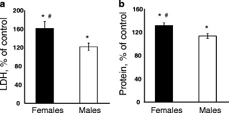
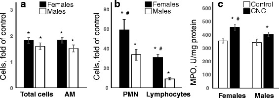
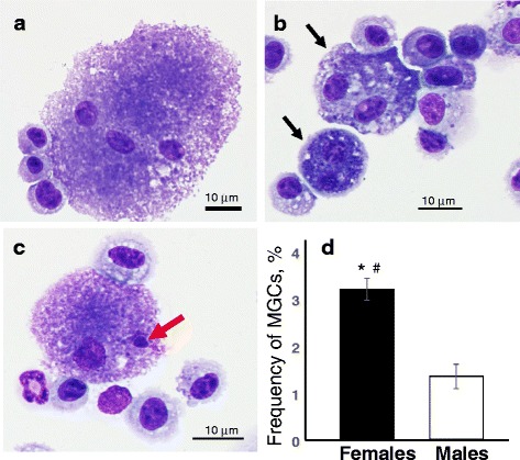
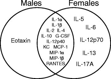
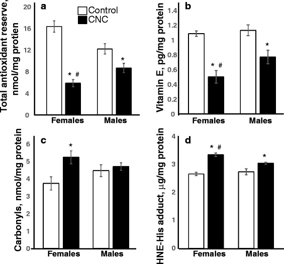
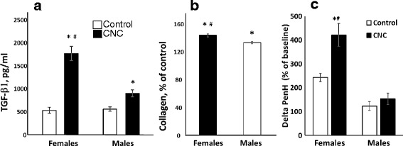
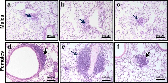
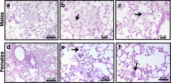
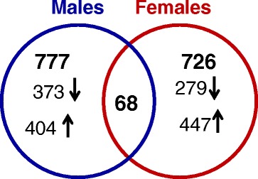
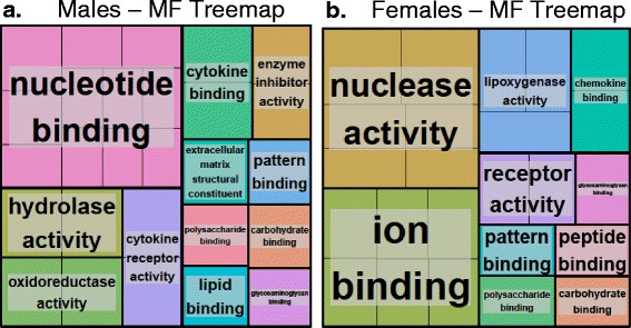
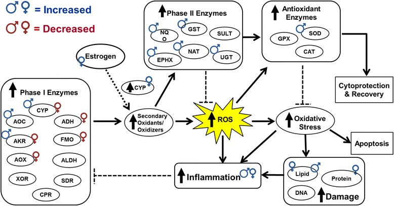
Results: Exposure to respirable CNC caused pulmonary inflammation and damage, induced oxidative stress, elevated TGF-β and collagen levels in lung, and impaired pulmonary functions. Notably, these effects were markedly more pronounced in females compared to male mice. Moreover, sex differences in responses to pulmonary exposure to CNC were also detected at the level of global mRNA expression as well as in inflammatory cytokine/chemokine activity.

Conclusions: Overall, our results indicate that there are considerable differences in responses to respirable CNC based on gender with a higher pulmonary toxicity observed in female mice.

Keywords: Cellulose; Gender differences; Inflammation; Oxidative stress; Pulmonary toxicity; mRNA expression.


Figures

Fig. 1
Fig. 1
Imaging of CNC using AFM height (a) and amplitude (b) modes, and SEM (c). The EDX spectrum is also shown (d), along with the calculated weight percentages (Wt%) and atomic percentages (At%). Silicon elemental contributions are ignored in the EDX analysis. Scale bars are 200 nm for a and b
Fig. 2
Fig. 2
Pulmonary tissue damage measured as LDH (a) and Protein (b) in BAL of C57BL/6 mice 3 months after repeated exposure to CNC (cumulative dose of 240 μg/mouse). Black columns – female mice exposed to CNC, open columns – male mice exposed to CNC. Mean absolute values of LDH in BAL from PBS-exposed control male and female mice were 53.8 ± 2.9 U/L and 52.3 ± 2.1 U/L, respectively. Mean absolute values of protein level in BAL from PBS-exposed control male and female mice were 0.238 ± 0.005 mg/ml and 0.224 ± 0.009 mg/ml, respectively. No significant differences were found between levels of LDH and protein measured in the BAL of controls from male and female mice. Mean ± SEM (n = 10 mice/group). *p < 0.05, vs control PBS-exposed mice, # p < 0.05, vs male mice exposed to CNC
Fig. 3
Fig. 3
Pulmonary inflammation evaluated by assessment of (a, b) cell profile from BAL of mice and (c) activity of myeloperoxidase (MPO) in lung homogenates of C57BL/6 mice 3 months after repeated pharyngeal aspiration with CNC (cumulative dose of 240 μg/mouse). a, b Black columns – female mice exposed to CNC, open columns – male mice exposed to CNC. Mean absolute values from PBS-exposed control female mice were (394.4 ± 28.6)x103, (373.8 ± 26.5)x103, 430.0 ± 214.0, and 396.0 ± 199.0 for total cells, alveolar macrophages, PMNs and lymphocytes, respectively. Mean absolute values from PBS-exposed control male mice were (343.6 ± 40.7)x103, (341.3 ± 40.2)x103, 645.3 ± 204.0, and 899.4 ± 218.0 for total cells, alveolar macrophages, PMNs and lymphocytes, respectively. No significant differences were found between number of total cells, alveolar macrophages, PMNs and lymphocytes in controls from female and male mice. Mean ± SEM (n = 10 mice/group). *p < 0.05, vs control PBS-exposed mice, # p < 0.05, vs male mice exposed to CNC. c Open columns – control mice, black columns –mice exposed to CNC. No significant differences were found between levels of MPO measured in the lung homogenates from females and males control mice. Mean ± SEM (n = 10 mice/group). *p < 0.05, vs control PBS-exposed mice, # p < 0.05, vs male mice exposed to CNC
Fig. 4
Fig. 4
Representative light micrographs of giant multi-nucleated (a-c) cells including bi-nucleated (b) and micro-nucleated (c) alveolar microphages from BAL fluids of female mice 3 month post repeated exposure with CNC (marked by arrows). Frequency of giant multi-nucleated cells (MGCs) in BAL fluids of female or male mice 3 month after the last exposure with CNC (d). Black columns – giant BAL cells from female mice exposed to CNC, open columns– giant BAL cells from male mice exposed to CNC. Mean ± SEM (n = 10 mice/group). *p < 0.05, vs control PBS-exposed mice, # p < 0.05, vs male mice exposed to CNC. Blind-coded slides were independently scored by two readers. A total of 2000 cells per sample were scored. No MGCs were found in control mice
Fig. 5
Fig. 5
A Venn diagram presenting differential cytokines/chemokines responses 3 month post repeated exposure of female and male C57BL/6 mice to CNC (cumulative dose of 240 mg/mouse). These measurements were performed using Bio-Rad 23-plex mouse assay kit, composed of a combination of pro- and anti- inflammatory cytokines with a subset of chemokine’s
Fig. 6
Fig. 6
Oxidative stress evaluated by measurements of a total antioxidant reserve, b vitamin E, c protein carbonyls and d HNE-His adduct in the lung homogenates of C57BL/6 mice 3 months after repeated exposure to CNC. Open columns – control mice, black columns – mice exposed to CNC. Mean ± SEM (n = 5-10 mice/group). *p < 0.05, vs control PBS-exposed mice, # p < 0.05, vs male mice exposed to CNC
Fig. 7
Fig. 7
Levels of TGF-β1 in the BAL (a), levels of collagen (b) and airway reactivity to direct stimulation of MCh (c) measured in the lung of female and male C57BL/6 mice 3 month post repeated exposure to CNC (cumulative dose of 240 μg/mouse). a & c Open columns – control mice, black columns – mice exposed to CNC. Mean ± SEM (n ≥ 5 mice/group). *p < 0.05, vs control PBS-exposed mice, # p < 0.05, vs male mice exposed to CNC. b Black columns – female mice exposed to CNC, open columns – male mice exposed to CNC. Mean absolute values of collagen in the lung of PBS-exposed control male and female mice were not significantly different (36.02 ± 1.90 μg/mg lung and 32.20 ± 1.50 μg/mg lung, respectively). Mean ± SEM (n = 10 mice/group). *p < 0.05, vs control PBS-exposed mice, # p < 0.05, vs male mice exposed to CNC
Fig. 8
Fig. 8
Light micrographs of H&E stained sections from lungs of female and male C57BL/6 mice 3 month post CNC repeated exposure showing chronic pulmonary inflammation: peribronchial (a & d), perivascular (b & e) and parenchymal (c & f)
Fig. 9
Fig. 9
Light micrographs of H&E stained sections from lungs of female and male C57BL/6 mice 3 month post CNC repeated exposure showing giant alveolar macrophages (a & d), bi- (b & e) and multi- nucleated (c & f) cells
Fig. 10
Fig. 10
Overlap of differentially expressed genes from both genders of C57BL/6 mice 3 months after repeated exposure to CNC
Fig. 11
Fig. 11
The enrichment of differentially expressed genes among molecular function category in mice exposed to CNC. Treemaps of DEGs in a males and b females generated using REVIGO. Each rectangle is a single cluster representative. The representatives are joined into ‘superclusters’ of loosely related terms, visualized with different colors. Size of the rectangles reflects either the p-value, or the frequency of the GO term in a given cluster. For more details please refer to methods section
Fig. 12
Fig. 12
Schematic representation of the detoxification/antioxidant mechanisms and biological effects associated with oxidative damage upon pulmonary exposure to CNC in male and female mice

Comment in

References

    1. Moon RJ, Martini A, Nairn J, Simonsen J, Youngblood J. Cellulose nanomaterials review: structure, properties and nanocomposites. Chem Soc Rev. 2011;40(7):3941–94. doi: 10.1039/c0cs00108b. - DOI - PubMed
    1. Postek MT, Moon RJ, Rudie A, Bilodeau M. Production and Applications of Cellulose Nanomaterials. Peachtree Corners, GA: TAPPI; 2013.
    1. Floros M, Hojabri L, Abraham E, Jose J, Thomas S, Pothan L, et al. Enhancement of thermal stability, strength and extensibility of lipid-based polyurethanes with cellulose-based nanofibers. Polym Degrad Stab. 2012;97(10):1970–8. doi: 10.1016/j.polymdegradstab.2012.02.016. - DOI
    1. H.P.S.A. K, A.H. B, A.F.I. Y. Green composites from sustainable cellulose nanofibrils: a review. Carbohyd Polym. 2012;87:963-79.
    1. Siqueira G, Bras J, Dufresne A. Cellulosic bionanocomposites: a review of preparation, properties and applications. Polym. 2010;2:728–65. doi: 10.3390/polym2040728. - DOI

Publication types

MeSH terms