Review
. 2016 Jul;229(1):32-62.
doi: 10.1111/joa.12473.

A brief history of topographical anatomy

Affiliations
Review

A brief history of topographical anatomy

Susan Standring. J Anat. 2016 Jul.

Abstract

This brief history of topographical anatomy begins with Egyptian medical papyri and the works known collectively as the Greco-Arabian canon, the time line then moves on to the excitement of discovery that characterised the Renaissance, the increasing regulatory and legislative frameworks introduced in the 18th and 19th centuries, and ends with a consideration of the impact of technology that epitomises the period from the late 19th century to the present day. This paper is based on a lecture I gave at the Winter Meeting of the Anatomical Society in Cambridge in December 2015, when I was awarded the Anatomical Society Medal.

Keywords: Galen; Harvey; Renaissance; Vesalius; dissection; legislation; technology.


Figures

Figure 1
Figure 1
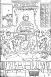
An academic dissection scene where the lector, seated in his high chair, reads the text, an ostensor demonstrates the anatomy and a barber‐surgeon (sector) carries out the dissection. From The Fasciculus Medicinae of Johannes de Ketham. Facsimile of the first, Venetian, edition of 1491. With introduction by Karl Sudhoff, translated and adapted by Charles Singer. R Lier & Co. Milan, 1924. Reproduced by kind permission of the President and Council of The Royal College of Surgeons of England.
Figure 2
Figure 2
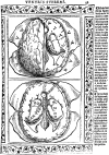
Brain dissected to reveal the ventricles at two levels, from Isagogae breves perlucide ac uberime in Anatomiam humani Corpori (Jacopo Berengario da Carpi, 1523). Reproduced by kind permission of the President and Council of The Royal College of Surgeons of England.
Figure 3
Figure 3
(A) Drawing of the nervous system from Mansur's text. Reproduced with permission from: Shoja MM, Tubbs RS (2007) The history of anatomy in Persia. J Anat 210, 359–378. (B) A pregnant woman. From The Fasciculus Medicinae of Johannes de Ketham. Facsimile of the first, Venetian, edition of 1491. With introduction by Karl Sudhoff, translated and adapted by Charles Singer. R Lier & Co Milan, 1924. Reproduced by kind permission of the President and Council of The Royal College of Surgeons of England.
Figure 4
Figure 4
The Battle of the Nude Men, Antonio Pollaiuolo, Florence ca. 1470. Reproduced by kind permission of The Metropolitan Museum of Art, New York.
Figure 5
Figure 5
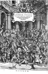
Title page. De humani corporis fabrica, Andreas Vesalius, Basel (1543). Reproduced by kind permission of the President and Council of The Royal College of Surgeons of England.
Figure 6
Figure 6
Three of the series of ‘muscle men’ from De humani corporis fabrica, Andreas Vesalius (1543). Reproduced by kind permission of the President and Council of The Royal College of Surgeons of England.
Figure 7
Figure 7
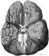
The base of a human brain (the first figure in Cerebri Anatome): the circle of Willis is illustrated. Nicolaus Steno (1638–1686) commented that …the best figures of the brain up to the present are those presented to us by Willis. Reproduced by kind permission of the President and Council of The Royal College of Surgeons of England.
Figure 8
Figure 8
Carl Manchot's vascular territories of the human integument (From Manchot C. Die Hautarterien des menschlichen Körpers. Leipzig: FCW Vogel; 1889). (A) Cutaneous vascular territories, ventral surface. (B) Cutaneous vascular territories, dorsal surface. Reproduced with permission from: Morris SF, Taylor GI (2012). Vascular territories. In: Plastic Surgery (ed. Neligan PC). Volume 1. Principles. Chapter 23, pp. 479–511. Saunders.
Figure 9
Figure 9
The angiosomes of the source arteries of the body. The angiosomes are: (1) thyroid; (2) facial; (3) buccal (internal maxillary); (4) ophthalmic; (5) superficial temporal; (6) occipital; (7) deep cervical; (8) transverse cervical; (9) acromiothoracic; (10) suprascapular; (11) posterior circumflex humeral; (12) circumflex scapular; (13) profunda brachii; (14) brachial; (15) ulnar; (16) radial; (17) posterior intercostals; (18) lumbar; (19) superior gluteal; (20) inferior gluteal; (21) profunda femoris; (22) popliteal; (22a) descending genicular (saphenous); (23) sural; (24) peroneal; (25) lateral plantar; (26) anterior tibial; (27) lateral femoral circumflex; (28) adductor (profunda); (29) medial plantar; (30) posterior tibial; (31) superficial femoral; (32) common femoral; (33) deep circumflex iliac; (34) deep inferior epigastric; (35) internal thoracic; (36) lateral thoracic; (37) thoracodorsal; (38) posterior interosseous; (39) anterior interosseous; and (40) internal pudendal. Reproduced with permission from: Morris SF, Taylor GI (2012). Vascular territories. In: Plastic Surgery (ed. Neligan PC). Volume 1. Principles. Chapter 23, pp. 479–511. Saunders.
Figure 10
Figure 10
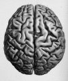
Superior view of the cerebral hemispheres with an accurate representation of the gyri and sulci, including variations in the precentral and postcentral gyri. Plate 11 in A.L Foville's Atlas published with Traité complet de l'anatomie, de la physiologie et de la pathologie du système nerveux cérébro‐spinal (1844). Reproduced by kind permission of the President and Council of The Royal College of Surgeons of England.

Similar articles

Cited by

References

    1. Abdel‐Halim RE (2001) Experimental medicine 1000 years ago. Urol Ann 3, 55–61. - PMC - PubMed
    1. Acierno LJ (1994) The History of Cardiology. London: The Parthenon Publishing Group; [Cited in Willerson JT, Teaff R (1996) Egyptian contributions to cardiovascular medicine. Tex Heart Inst J 23, 191–200]. - PMC - PubMed
    1. Afif A, Mertens P (2010) Description of sulcal organization of the insular cortex. Surg Radiol Anat 32, 491–498. - PubMed
    1. Agrawal A, Kapfhammer JP, Kress A, et al. (2011) Josef Klingler's models of white matter tracts: influences on neuroanatomy, neurosurgery, and neuroimaging. Neurosurgery 69, 238–252. - PubMed
    1. Aird WC (2011) Discovery of the cardiovascular system: from Galen to William Harvey. J Thromb Haemost 9(Suppl. 1), 118–129. - PubMed

LinkOut - more resources