

3D-Microarchitectural patterns of Hyperostosis frontalis interna: a micro-computed tomography study in aged women
- PMID: 27279170
- PMCID: PMC5055089
- DOI: 10.1111/joa.12506
3D-Microarchitectural patterns of Hyperostosis frontalis interna: a micro-computed tomography study in aged women
Abstract
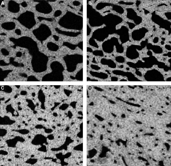
Although seen frequently during dissections and autopsies, Hyperostosis frontalis interna (HFI) - a morphological pattern of the frontal bone thickening - is often ignored and its nature and development are not yet understood sufficiently. Current macroscopic classification defines four grades/stages of HFI based on the morphological appearance and size of the affected area; however, it is unclear if these stages also depict the successive phases in the HFI development. Here we assessed 3D-microarchitecture of the frontal bone in women with various degrees of HFI expression and in an age- and sex-matched control group, hypothesizing that the bone microarchitecture bears imprints of the pathogenesis of HFI and may clarify the phases of its development. Frontal bone samples were collected during routine autopsies from 20 women with HFI (age: 69.9 ± 11.1 years) and 14 women without HFI (age: 74.1 ± 9.7 years). We classified the HFI samples into four groups, each group demonstrating different macroscopic type or stage of HFI. All samples were scanned by micro-computed tomography to evaluate 3D bone microarchitecture in the following regions of interest: total sample, outer table, diploe and inner table. Our results revealed that, compared to the control group, the women with HFI showed a significantly increased bone volume fraction in the region of diploe, along with significantly thicker and more plate-like shaped trabeculae and reduced trabecular separation and connectivity density. Moreover, the inner table of the frontal bone in women with HFI displayed significantly increased total porosity and mean pore diameter compared to controls. Microstructural reorganization of the frontal bone in women with HFI was also reflected in significantly higher porosity and lower bone volume fraction in the inner vs. outer table due to an increased number of pores larger than 100 μm. The individual comparisons between the control group and different macroscopic stages of HFI revealed significant differences only between the control group and the morphologically most pronounced type of HFI. Our microarchitectural findings demonstrated clear differences between the HFI and the control group in the region of diploe and the inner table. Macroscopic grades of HFI could not be distinguished at the level of bone microarchitecture and their consecutive nature cannot be supported. Rather, our study suggests that only two different types of HFI (moderate and severe HFI) have microstructural justification and should be considered further. It is essential to record HFI systematically in human postmortem subjects to provide more data on the mechanisms of its development.
Keywords: frontal bone; hyperostosis; micro-architecture; women.
© 2016 Anatomical Society.
Figures


References
-
- Attanasio F, Granziera S, Giantin V, et al. (2013) Full penetrance of Morgagni‐Stewart‐Morel syndrome in 75‐years old women: case report and review of the literature. J Clin Endocrinol Metab 98, 453–457. - PubMed
-
- Brandi ML, Collin‐Osdoby P (2006) Vascular biology and the skeleton. J Bone Miner Res 21, 183–192. - PubMed
-
- Cetiner Batun G, Yuruyen M, Vatankulu B, et al. (2015) Hyperostosis frontalis interna presenting as depression and parkinsonism in an older woman. Psychogeriatrics doi: 10.1111/psyg.12166. [Epub ahead of print]. - DOI - PubMed
-
- Devriendt W, Piercecchi‐Marti MD, Adalian P, et al. (2005) Hyperostosis frontalis interna: forensic issues. J Forensic Sci 50, 143–146. - PubMed
-
- Djonic D, Bracanovic D, Rakocevic Z, et al. (2016) Hyperostosis frontalis interna in postmenopausal women – possible relation to osteoporosis. Women Health 19, 1–14. - PubMed
Publication types
MeSH terms
LinkOut - more resources
Full Text Sources
Other Literature Sources
