. 2016 Jun 9:6:27641.
doi: 10.1038/srep27641.

MicroRNA-320a promotes 5-FU resistance in human pancreatic cancer cells

Affiliations

MicroRNA-320a promotes 5-FU resistance in human pancreatic cancer cells

Weibin Wang et al. Sci Rep. .

Abstract

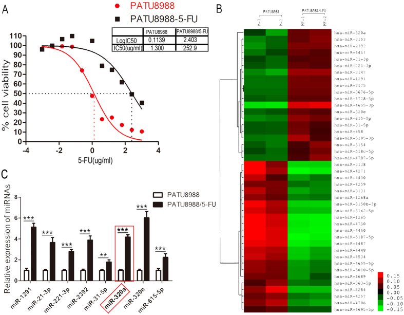
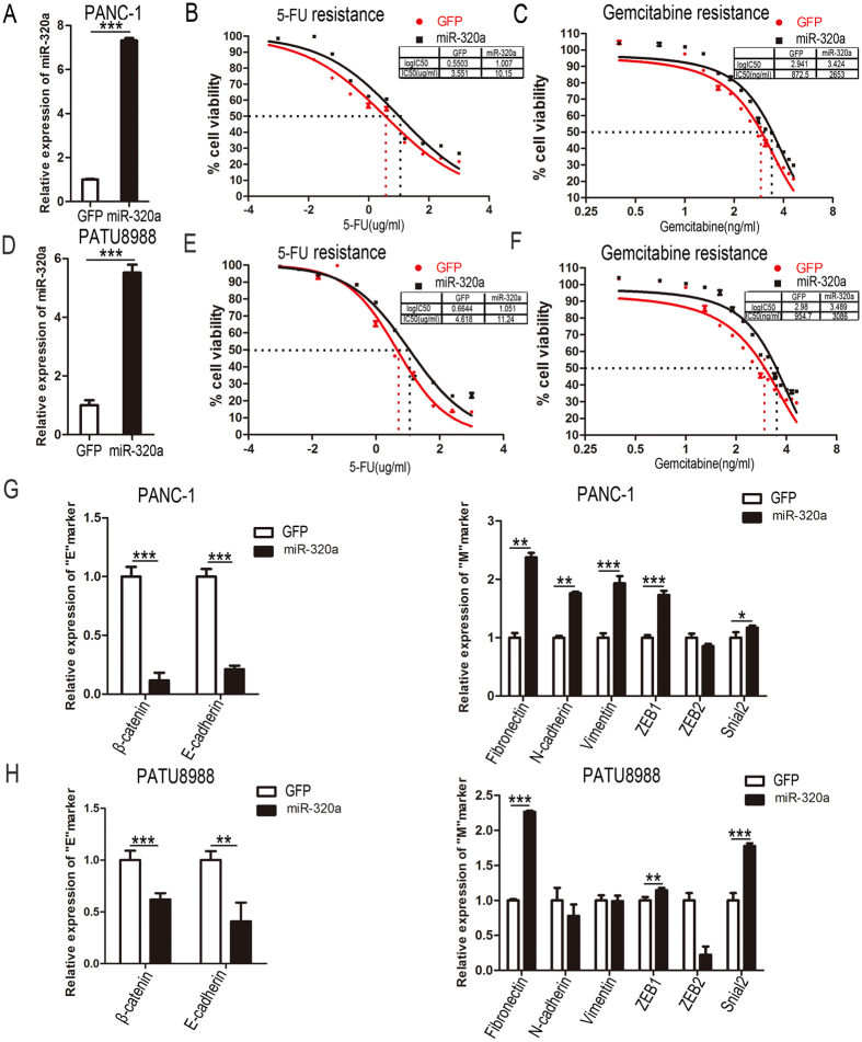
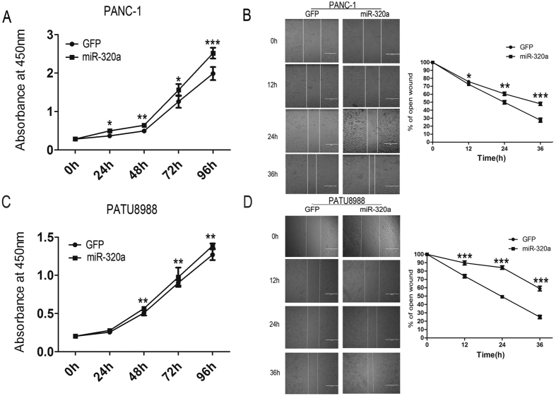
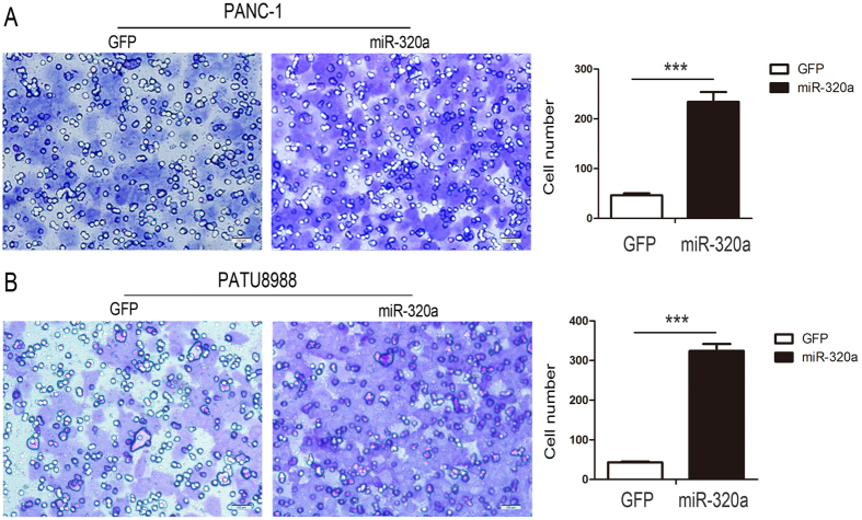
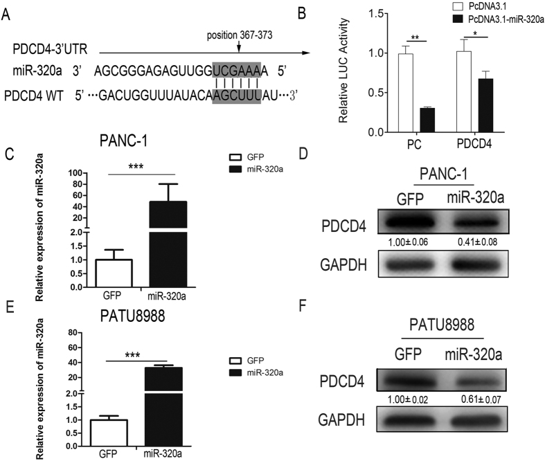
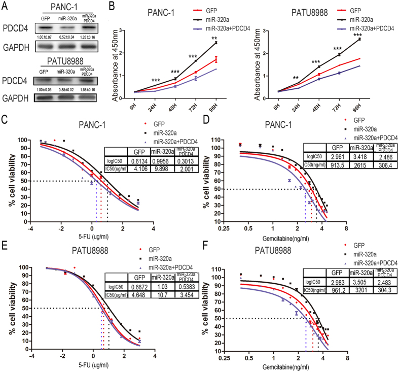
The drug-resistance of pancreatic cancer cells results in poor therapeutic effect. To predict the therapeutic effect of the chemotherapy drugs to specific patients and to reverse the resistance of pancreatic cancer cells are critical for chemotherapy of pancreatic cancer. MicroRNAs (miRNAs) have been reported to play important roles in the genesis of drug-resistance of various cancer types. There are also many advantages of miRNAs in diagnosis and therapy of disease. Although several miRNAs regulating 5-Fluorouracil (5-FU) resistance in human pancreatic cancer have been reported, the detailed molecular mechanism remains to be determined. In this study, we found that miR-320a was significantly up-regulated in 5-FU resistant pancreatic cancer cells. Over-expression of miR-320a strongly contributed to pathogenesis of pancreatic cancer, which was represented by the increased proliferation, invasion, metastasis, drug-resistance characteristics and the epithelial-to-mesenchymal transition. Furthermore, we demonstrated that miR-320a was able to bind to 3'UTR of PDCD4 mRNA, and mediated its down-regulation in 5-FU resistance of human pancreatic cancer cells. Whereas restoration of PDCD4 expression could partially attenuate the function of miR-320a in pancreatic cancer. Taken together, our study demonstrated that miR-320a played important role in regulating 5-FU resistance by targeting PDCD4 and might be developed as new therapeutic target for pancreatic cancer.


Figures

Figure 1
Figure 1. Up-regulation of miR-320a in PATU8988/5-FU cells.
(A) Representative curves of growth-inhibitory effects in 5-FU resistant PATU8988/5-FU and PATU8988 cells. IC50, inhibitory concentration 50. (B) Microarray of miRNA expression profile of PATU8988/5-FU and PATU8988 cells. Each sample is biologically duplicated. (C) Up-regulated microRNAs between 5-FU resistant PATU8988/5-FU cells and its parental PATU8988 cells. All data are presented as the means ± SD. Student’s t test (two-tailed) was performed to analyze data from the experiments in triplicate. P values < 0.05 were considered significant, as indicated by the asterisks (**p < 0.01, ***p < 0.001).
Figure 2
Figure 2. Over-expression of miR-320a induces resistance to 5-FU in pancreatic cancer cells.
(A) RT-PCR results showed the expression of miR-320a in PANC-1 cells after lentivirus infection with lenti_GFP and lenti_miR-320a. (B) Representative curves of growth-inhibitory effects of 5-FU in PANC-1 cells which infected with lenti_miR-320a and lenti_GFP. (C) Representative curves of growth-inhibitory effects of gemcitabine in PANC-1 cells which infected with lenti_miR-320a and lenti_GFP. (D) RT-PCR results of the expression of miR-320a in PATU8988 cells after lentivirus infection with lenti_GFP and lenti_miR-320a. (E) Representative curves of growth-inhibitory effects of 5-FU in PATU8988 cells after infecting with lenti_miR-320a and lenti_GFP. (F) Representative curves of growth-inhibitory effects of gemcitabine in PATU8988cells which infected with lenti_miR-320a and lenti_GFP. (G,H) Real-time PCR assay was conducted to detect the expression of “EMT” markers in PANC-1 cells and PATU8988 cells. Left panel: the expression of β-cadherin, E-cadherin, Right panel: the expression of Fibronectin N-cadherin, Vimentin, ZEB1, ZEB2 and Snail in PANC-1 cells. All data are presented as the means ± SD. Student’s t test (two-tailed) was performed to analyze data from the experiments in triplicate. P values < 0.05 were considered significant, as indicated by the asterisks (*p < 0.05, **p < 0.01, ***p < 0.001).
Figure 3
Figure 3. Over-expression of miR-320a promotes pancreatic cancer cell proliferation and migration.
(A) Proliferation assays by cck-8 at 0, 24, 48, 72 and 96 h, after infecting with lenti_miR-320a or lenti_GFP in PANC-1 cells. (B) Left panel: PANC-1 cells were infected with lenti_miR-320a or lenti_GFP for 36 h, and wounds were made. Bar, 100 uM. Right panel: The relative ratio of wound closure per field is shown at 0, 12, 24 and 36 h. (C) Proliferation assays by cck-8 at 0, 24, 48, 72 and 96 h, after infecting with lenti_miR-320a or lenti_GFP in PATU8988 cells. (D) Left panel: PATU8988 cells were infected with lenti_miR-320a or lenti_GFP for 36 h, and wounds were made. Bar, 100 uM. Right panel: The relative ratio of wound opening per field is shown at 0, 12, 24 and 36 h. All data are presented as the means ± SD. Student’s t test (two-tailed) was performed to analyze data from the experiments in triplicate. P values < 0.05 were considered significant, as indicated by the asterisks (*p < 0.05, **p < 0.01, ***p < 0.001).
Figure 4
Figure 4. Over-expression of miR-320a promotes pancreatic cancer Cell invasion.
(A) PANC-1 cells were infected with lenti_miR-320a or lenti_GFP for 24 hours, and transwell invasion assay were performed. The invasive cells per field is shown. (B) PATU8988 cells were infected with lenti_miR-320a or lenti_GFP for 24 hours, and transwell invasion assay were performed. The number of invasive cells per field is shown. All data are presented as the means ± SD. ***p < 0.001.
Figure 5
Figure 5. PDCD4 is a direct target of miR-320a in pancreatic cancer cells.
(A)The prediction of the binding between miR-320a and PDCD4 by Pictar. (B) Relative luciferase activity of the indicated PDCD4 reporter constructs in 293T cells. Error bars presented standard deviation obtained from three independent experiments. (C) RT-PCR showed the expression of miR-320a in PANC-1 cells after lentivirus infection with lenti_GFP and lenti_miR-320a. (D) Western blot analysis showed PDCD4 protein levels in PANC-1 after miR-320a over expression in pancreatic cancer cells. (E) RT-PCR results about the expression of miR-320a in PATU8988 cells after lentivirus infection with lenti_GFP and lenti_miR-320a. (F) Western bot analysis showed PDCD4 protein levels in PATU8988 after miR-320a over expression in pancreatic cancer cells. All data are presented as the means ± SD. Student’s t test (two-tailed) was performed to analyze data from the experiments in triplicate. P values < 0.05 were considered significant, as indicated by the asterisks (*p < 0.05, **p < 0.01, ***p < 0.001).
Figure 6
Figure 6. PDCD4 could rescue the increased proliferation rate and drug resistance induced by miR-320a in pancreatic cancer cells.
(A) Western blot analysis of PDCD4 in pancreatic cancer cells with miR-320a over-expression transfected with PDCD4. (B) Proliferation assays by cck-8 at 0, 24, 48, 72 and 96 h after transfected with PDCD4 in PANC-1 and PATU8988 cells. (C) Representative curves of growth-inhibitory effects of 5-FU in PANC-1 cells. (D) Representative curves of growth-inhibitory effects of gemcitabine in PANC-1 cells. (E) Representative curves of growth-inhibitory effects of 5-FU in PATU8988 cells. (F) Representative curves of growth-inhibitory effects of gemcitabine in PATU8988 cells. All data are presented as the means ± SD. Student’s t test (two-tailed) was performed to analyze data from the experiments in triplicate. P values < 0.05 were considered significant, as indicated by the asterisks (**p < 0.01, ***p < 0.001).

References

    1. Siegel R., Naishadham D. & Jemal A. Cancer statistics, 2013. CA Cancer J Clin 63, 11–30, doi: 10.3322/caac.21166 (2013). - DOI - PubMed
    1. Jemal A., Siegel R., Xu J. & Ward E. Cancer statistics, 2010. CA Cancer J Clin 60, 277–300, doi: 10.3322/caac.20073 (2010). - DOI - PubMed
    1. Chen W. et al.. Report of incidence and mortality in China cancer registries, 2009. Chin J Cancer Res 25, 10–21, doi: 10.3978/j.issn.1000-9604.2012.12.04 (2013). - DOI - PMC - PubMed
    1. Gottesman M. M. Mechanisms of cancer drug resistance. Annu Rev Med 53, 615–627, doi: 10.1146/annurev.med.53.082901. (2002). - DOI - PubMed
    1. Hermann P. C. et al.. Distinct populations of cancer stem cells determine tumor growth and metastatic activity in human pancreatic cancer. Cell Stem Cell 1, 313-–323., doi: 10.1016/j.stem.2007.06.002 (2007). - DOI - PubMed

Publication types

MeSH terms