. 2016 Jul 27;90(16):7529-7540.
doi: 10.1128/JVI.00656-16. Print 2016 Aug 15.

Arabidopsis RNA Polymerases IV and V Are Required To Establish H3K9 Methylation, but Not Cytosine Methylation, on Geminivirus Chromatin

Affiliations

Arabidopsis RNA Polymerases IV and V Are Required To Establish H3K9 Methylation, but Not Cytosine Methylation, on Geminivirus Chromatin

Jamie N Jackel et al. J Virol. .

Abstract

In plants, RNA-directed DNA methylation (RdDM) employs small RNAs to target enzymes that methylate cytosine residues. Cytosine methylation and dimethylation of histone 3 lysine 9 (H3K9me2) are often linked. Together they condition an epigenetic defense that results in chromatin compaction and transcriptional silencing of transposons and viral chromatin. Canonical RdDM (Pol IV-RdDM), involving RNA polymerases IV and V (Pol IV and Pol V), was believed to be necessary to establish cytosine methylation, which in turn could recruit H3K9 methyltransferases. However, recent studies have revealed that a pathway involving Pol II and RNA-dependent RNA polymerase 6 (RDR6) (RDR6-RdDM) is likely responsible for establishing cytosine methylation at naive loci, while Pol IV-RdDM acts to reinforce and maintain it. We used the geminivirus Beet curly top virus (BCTV) as a model to examine the roles of Pol IV and Pol V in establishing repressive viral chromatin methylation. As geminivirus chromatin is formed de novo in infected cells, these viruses are unique models for processes involved in the establishment of epigenetic marks. We confirm that Pol IV and Pol V are not needed to establish viral DNA methylation but are essential for its amplification. Remarkably, however, both Pol IV and Pol V are required for deposition of H3K9me2 on viral chromatin. Our findings suggest that cytosine methylation alone is not sufficient to trigger de novo deposition of H3K9me2 and further that Pol IV-RdDM is responsible for recruiting H3K9 methyltransferases to viral chromatin.

Importance: In plants, RNA-directed DNA methylation (RdDM) uses small RNAs to target cytosine methylation, which is often linked to H3K9me2. These epigenetic marks silence transposable elements and DNA virus genomes, but how they are established is not well understood. Canonical RdDM, involving Pol IV and Pol V, was thought to establish cytosine methylation that in turn could recruit H3K9 methyltransferases, but recent studies compel a reevaluation of this view. We used BCTV to investigate the roles of Pol IV and Pol V in chromatin methylation. We found that both are needed to amplify, but not to establish, DNA methylation. However, both are required for deposition of H3K9me2. Our findings suggest that cytosine methylation is not sufficient to recruit H3K9 methyltransferases to naive viral chromatin and further that Pol IV-RdDM is responsible.


Figures

FIG 1
FIG 1
Plants deficient for Pol IV, Pol V, and CLSY1 are hypersusceptible to geminivirus infection. Photographs taken under a dissecting microscope show floral heads from infected wild-type (Col-0), clsy1, pol IV, pol V, and pol IV/V plants. Primary shoots infected with BCTV (top row) were photographed ∼21 days postinoculation, and photographs of secondary infected shoots infected with BCTV L2 (bottom row) were taken ∼14 days after harvest of primary tissue (∼35 days postinoculation). Photographs are representative of 32 plants per treatment. Note increased floral deformation in mutant compared to wild-type plants during primary infection. During secondary infection (recovery assay), the absence of symptoms indicates recovery in wild-type plants, while pol V and pol IV/V mutants do not recover and show severe symptoms. The pol IV and clsy1 mutants exhibit delayed recovery. Nonrecovered (NR) and recovered (R) tissue was harvested as indicated (white lines).
FIG 2
FIG 2
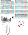
Pol IV and Pol V are not required to establish viral DNA methylation. (A) Methylation status of the BCTV L2 IR from secondary tissue of infected wild-type and mutant plants. Samples from recovered (R) and nonrecovered (NR) regions of shoots from clsy1 and pol IV mutants were separately analyzed (Fig. 1). Samples consisted of tissue pooled from three plants. DNA was treated with bisulfite, and PCR was performed to amplify the viral strand. PCR products were cloned and sequenced. Rows depict individual clones (12 per treatment) organized from most to least methylated. Each circle represents a cytosine in the IR (42 total) with CG (10) in red, CHG (7) in blue, and CHH (25) in green. Filled circles indicate a methylated cytosine. (B) Histograms show the percentage of methylated cytosine residues in different sequence contexts. Total methylation is indicated in gray. Bars indicate Wilson score interval 95% confidence limits. (C) Compilation of bisulfite data from wild-type and mutant plants. Clones from secondary tissues of BCTV L2-infected plants classified as recovered (n = 93) or nonrecovered (n = 90) were placed in separate groups (see the text). Asterisks indicate mean cytosine methylation levels are significantly different (P < 0.001), as determined by Student's t test. (D) The diagram depicts the dsDNA replicative form of BCTV. Genes are shown as gray arrows, with the L2 silencing suppressor in red. The replication initiation site within the IR is marked with an asterisk, and Pol II transcription starts are indicated by right-angle arrows.
FIG 3
FIG 3
Pol II, Pol IV, and Pol V associate with the BCTV genome. (A) Representative ChIP experiments with extracts from infected, transgenic plants expressing FLAG-Pol II, Pol IV, or Pol V subunits are shown. Extracts were obtained from BCTV-infected primary tissue (blue bars) and BCTV L2-infected secondary tissue (red bars), and in all cases samples consisted of pooled tissue from at least three plants. DNA precipitated with FLAG antibody was analyzed using qPCR performed in triplicate with at least three independent samples (three biological replicates with three technical replicates each). Primer sets to amplify the BCTV IR and the CP CDS (CP2) were employed, as were control primers for actin and IGN5. IgG was a background control. The graphs depict signal as a percentage of input. Bars indicate standard errors. (B) Methylation of the CP CDS in nonrecovered and recovered tissues. Cytosine methylation in the CP CDS was assessed in primary (1°) tissue from wild-type and pol IV/V plants infected with BCTV or BCTV L2 and in BCTV L2-infected secondary (2°) tissue of recovered wild-type plants and nonrecovered pol IV/V plants. Infected inflorescence tissue was harvested from a pool of at least three plants, and DNA was isolated and treated with sodium bisulfite to convert unmethylated cytosines to uracil. PCR with primers designed to amplify CP2 (Table 1) was performed, and products were cloned and sequenced. The region examined contains 46 cytosines: 7 CG, 11 CHG, and 28 CHH. Twelve clones were examined per treatment. Histograms show total cytosine methylation levels (gray bars) as well as methylation in different sequence contexts (CG in red, CNG in blue, and CHH in green). Bars represent Wilson score interval 95% confidence limits.
FIG 4
FIG 4
ChIP analysis (sqPCR) of Pol II, Pol IV, and Pol V association with the BCTV genome. Results of ChIP experiments with extracts from infected, transgenic plants expressing FLAG-Pol II, Pol IV, or Pol V subunits are shown. (A) Extracts were obtained from BCTV-infected primary tissue. (B) Extracts were obtained from BCTV L2-infected secondary tissue. Using DNA precipitated with FLAG antibody, PCR products (serial 2-fold dilutions shown) were generated with IR primers and CP1 primers or with IGN5 and actin primers (controls). In all cases samples consisted of tissue pooled from at least three plants. The experiments shown are representative of at least three independent trials. IgG was a background control.
FIG 5
FIG 5
Pol IV and Pol V are not required to generate BCTV-specific siRNAs. (A) Northern blot representative of three independent experiments shows virus-specific small RNAs from BCTV-infected primary tissue of wild-type (Col-0), clsy1, pol IV, pol V, and pol IV/V plants. In each case samples consisted of inflorescence tissue pooled from at least three infected plants. The probes used were specific for the BCTV IR. siRNA levels were normalized to rRNA loading controls visualized by ethidium bromide staining. Note that the lanes shown were from the same gel spliced to remove intervening samples. (B) The graph compares relative siRNA levels in wild-type plants and plants harboring the indicated mutations, normalized to rRNA loading controls. Mean levels of total, 24-nt, and 21/22-nt siRNAs observed in wild-type Col-0 plants were set to 1. Data were compiled from three independent experiments. Bars indicate standard errors.
FIG 6
FIG 6
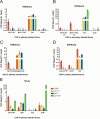
Pol IV and Pol V are required for deposition of H3K9me2 on viral chromatin. Representative ChIP experiments are shown. (A to E) Extracts were prepared from wild-type (Col-0, red bars), pol IV (yellow bars), pol V (blue bars), or pol IV/V (green bars in A, B, and E) plants inoculated with BCTV or CaLCuV (primary infected tissue) or with BCTV L2 (secondary infected tissue). In all cases, samples consisted of inflorescence tissue pooled from at least three infected plants. ChIP was performed using antibody against H3K9me2 or H3 acetylated at lysine 9 and 14 (H3-Ac) as indicated, and qPCR was carried out in triplicate with primer sets to amplify the viral IR or the CP CDS (CP2) (Table 1). Transposon Ta3 or Athila6A was a positive control for H3K9me2, while actin was a positive control for H3-Ac. The graphs show ChIP signal as a percentage of the input. Graphs in panels A, B, and E are representative of three biological replicates with three technical replicates each (with the exception of BCTV L2 pol V), while panels C and D are representative of two biological replicates with three technical replicates each. The experiment shown in panel C is a partial repeat of panel A but using the Athila6A LTR instead of Ta3 as a positive control. Bars indicate standard errors.

References

    1. Law JA, Jacobsen SE. 2010. Establishing, maintaining, and modifying DNA methylation patterns in plants and animals. Nat Rev Genet 11:204–220. doi: 10.1038/nrg2719. - DOI - PMC - PubMed
    1. Matzke MA, Mosher RA. 2014. RNA-directed DNA methylation: an epigenetic pathway of increasing complexity. Nat Rev Genet 15:394–408. doi: 10.1038/nrg3683. - DOI - PubMed
    1. Herr AJ, Jensen MB, Dalmay T, Baulcombe DC. 2005. RNA polymerase IV directs silencing of endogenous DNA. Science 308:118–120. doi: 10.1126/science.1106910. - DOI - PubMed
    1. Kanno T, Huettel B, Mette MF, Aufsatz W, Jaligot E, Daxinger Kreil LDP, Matzke M, Matzke AJ. 2005. Atypical RNA polymerase subunits required for RNA-directed DNA methylation. Nat Genet 37:761–765. doi: 10.1038/ng1580. - DOI - PubMed
    1. Onodera Y, Haag JR, Ream T, Nunes PC, Pontes O, Pikaard CS. 2005. Plant nuclear RNA polymerase IV mediates siRNA and DNA methylation-dependent heterochromatin formation. Cell 120:613–622. doi: 10.1016/j.cell.2005.02.007. - DOI - PubMed

Publication types

LinkOut - more resources