

Three-dimensional reconstruction and morphological characterization of pituitary macroadenomas
- PMID: 27279851
- PMCID: PMC4889693
- DOI: 10.5114/aoms.2016.59932
Three-dimensional reconstruction and morphological characterization of pituitary macroadenomas
Abstract
Introduction: The aim was to investigate the relationship between the tumor (clinicopathologic and radiological) characteristics and the morphological parameters of pituitary macroadenoma or giant adenoma patients using a three-dimensional (3D) reconstructed model.
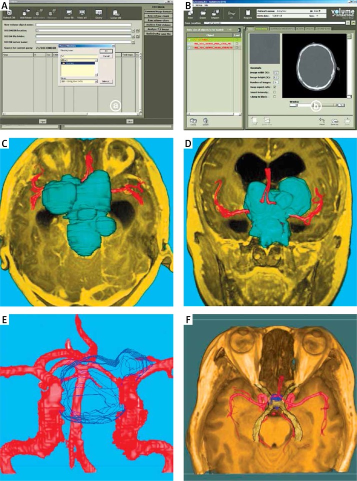
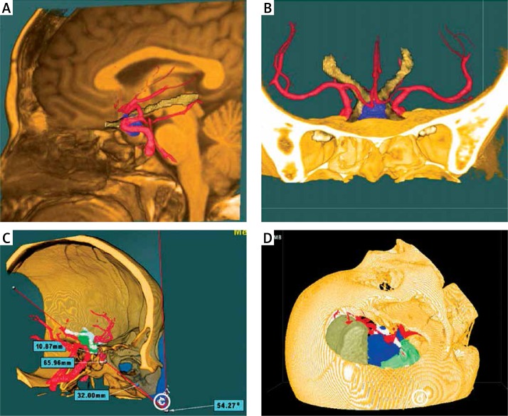
Material and methods: Magnetic resoanance imaging (MRI) was performed preoperatively; tumor grade was determined by the Knosp-Steiner classification and tumor morphology by the SIPAP classification. Pituitary adenomas and adjacent structures were reconstructed three-dimensionally by volume rendering.
Results: Fifty-two and 6 patients underwent surgery via the transnasal transsphenoidal or pterional approach, respectively. Knosp-Steiner grades I to IV adenomas were observed in 5.2%, 25.9%, 22.4% and 46.6% of the patients, respectively. The 3D model was reconstructed in all cases with superb delineation of tumor morphology and the spatial relationship between the tumor and adjacent tissues. Pituitary adenomas were categorized into intrasellar (13.8%), suprasellar (20.7%), infrasellar (17.2%), and lobulated adenomas (48.3%). Suprasellar adenomas had the smallest (2.27 ±3.22 cm(3)) and lobulated adenomas the largest volume (24.61 ±30.50 cm(3)). Intrasellar adenomas were all functioning, while 75%, 60% and 60.7%, respectively, of suprasellar, infrasellar and lobulated adenomas were nonfunctioning, with a significant association between tumor morphology and secretory function (p = 0.005).
Conclusions: Three-dimensional reconstruction of pituitary macroadenomas offers a simplified morphological classification of pituitary adenomas and may be helpful for neurosurgeons to categorize and characterize pituitary adenomas.
Keywords: 3D reconstruction; SIPAP classification; morphology; pituitary macroadenomas; volume rendering.
Figures

References
-
- Asa SL, Ezzat S. The pathogenesis of pituitary tumours. Nat Rev Cancer. 2002;2:836–49. - PubMed
-
- Scheithauer BW, Kovacs KT, Laws ER, Jr, Randall RV. Pathology of invasive pituitary tumors with special reference to functional classification. J Neurosurg. 1986;65:733–44. - PubMed
-
- Colao A, Grasso LF, Pivonello R, Lombardi G. Therapy of aggressive pituitary tumors. Expert Opin Pharmacother. 2011;12:1561–70. - PubMed
-
- Ferroli P, Tringale G, Acerbi F, Aquino D, Franzini A, Brogg G. Brain surgery in a stereoscopic virtual reality environment: a single institution's experience with 100 cases. Neurosurgery. 2010;67(ONS Suppl. 1):ons 79–84. - PubMed
-
- Hao B, Zhou X, Jin A, et al. Preliminary exploration of three dimensional reconstruction of MRI images of human subthalamic nucleus. Chin J Neurosurg. 2010;26:548–51.
LinkOut - more resources
Full Text Sources
Other Literature Sources