Case Reports
. 2016 Jun;6(3):262-6.
doi: 10.21037/cdt.2015.11.05.

Conus artery occlusion causing isolated right ventricular outflow tract infarction: novel application of cardiac magnetic resonance in anterior STEMI

Affiliations
Case Reports

Conus artery occlusion causing isolated right ventricular outflow tract infarction: novel application of cardiac magnetic resonance in anterior STEMI

Melissa Lyle et al. Cardiovasc Diagn Ther. 2016 Jun.

Abstract

Acute ST elevation in the anterior precordial leads typically suggests an anteroseptal infarction due to left anterior descending coronary artery obstruction, but the differential can be broad. Conus branch artery occlusion is a potentially overlooked cause of anteroseptal ST elevation myocardial infraction. Cardiac magnetic resonance (CMR) imaging is an emerging technology which can differentiate the etiology of anterior ST elevation in patients with no apparent coronary abnormalities on coronary angiography and normal echocardiography.

Keywords: Conus artery; bicuspid aortic valve; magnetic resonance imaging (MRI); myocardial infarction.


Conflict of interest statement

Conflicts of Interest: The authors have no conflicts of interest to declare.

Figures

Figure 1
Figure 1
Coronal T2 weighted and axial ADC map diffusion weighted images of the brain revealing signal in the inferolateral right temporal lobe, indicative of a chronic middle cerebral artery (MCA) territory infarction.
Figure 2
Figure 2
Parasternal long axis view of the bicuspid aortic valve demonstrating the mobile echo densities.
Figure 3
Figure 3
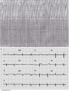
Electrocardiogram illustrates ST segment elevations in leads V1-V3.
Figure 4
Figure 4
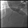
Aortogram used to visualize the right coronary artery (RCA). A conus branch was not visualized on the aortogram or injection of the RCA (not shown).
Figure 5
Figure 5
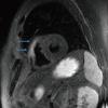
T2 weighted triple inversion recovery fast spin echo magnetic resonance imaging (MRI) sequence illustrating right ventricular outflow tract (RVOT) myocardial edema.
Figure 6
Figure 6
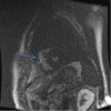
Still frame from dynamic first pass perfusion imaging (FGRET). Blue arrows highlight lack of perfusion in the right ventricular outflow tract (RVOT).
Figure 7
Figure 7
Late gadolinium enhancement of the right ventricular outflow tract (RVOT). Blue arrows demonstrate subtle enhancement of thin right ventricular (RV) myocardium.

References

    1. Porter A, Herz I, Strasberg B. Isolated right ventricular infarction presenting as anterior wall myocardial infarction on electrocardiography. Clin Cardiol 1997;20:971-3. 10.1002/clc.4960201115 - DOI - PMC - PubMed
    1. Levin DC, Beckmann CF, Garnic JD, et al. Frequency and clinical significance of failure to visualize the conus artery during coronary arteriography. Circulation 1981;63:833-7. 10.1161/01.CIR.63.4.833 - DOI - PubMed
    1. Muhammad KI, Kapadia SR. Anterior ST-segment elevation with right coronary artery occlusion: a unique case of isolated right ventricular infarction. Angiology 2008;59:622-4. 10.1177/0003319707305464 - DOI - PubMed
    1. Fabris E, Morocutti G, Sinagra G, et al. Uncommon cause of ST-segment elevation in V1-V3: incremental value of cardiac magnetic resonance imaging. Clin Res Cardiol 2014;103:825-8. 10.1007/s00392-014-0715-1 - DOI - PubMed
    1. Erol C, Seker M. The prevalence of coronary artery variations on coronary computed tomography angiography. Acta Radiol 2012;53:278-84. 10.1258/ar.2011.110394 - DOI - PubMed

Publication types

LinkOut - more resources