. 2016 Aug 1;27(15):2360-7.
doi: 10.1091/mbc.E16-01-0032. Epub 2016 Jun 8.

Hyperactivation of ATM upon DNA-PKcs inhibition modulates p53 dynamics and cell fate in response to DNA damage

Affiliations

Hyperactivation of ATM upon DNA-PKcs inhibition modulates p53 dynamics and cell fate in response to DNA damage

Ana Finzel et al. Mol Biol Cell. .

Abstract

A functional DNA damage response is essential for maintaining genome integrity in the presence of DNA double-strand breaks. It is mainly coordinated by the kinases ATM, ATR, and DNA-PKcs, which control the repair of broken DNA strands and relay the damage signal to the tumor suppressor p53 to induce cell cycle arrest, apoptosis, or senescence. Although many functions of the individual kinases have been identified, it remains unclear how they act in concert to ensure faithful processing of the damage signal. Using specific inhibitors and quantitative analysis at the single-cell level, we systematically characterize the contribution of each kinase for regulating p53 activity. Our results reveal a new regulatory interplay in which loss of DNA-PKcs function leads to hyperactivation of ATM and amplification of the p53 response, sensitizing cells for damage-induced senescence. This interplay determines the outcome of treatment regimens combining irradiation with DNA-PKcs inhibitors in a p53-dependent manner.


Figures

FIGURE 1:
FIGURE 1:
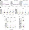
ATM, ATR, and DNA-PK control the p53 response to DSBs. (A) Schematic overview of the p53 network in response to DSBs. (B) Time-lapse microscopy images of A549 cells expressing p53-YFP after treatment with 10-Gy γ-IR. (C) Individual A549 cells were tracked, and the p53 average nuclear fluorescence intensity was measured in cells untreated or treated with a mix of ATMi, ATRi, and DNA-PKi. (D) Quantification of the p53 average nuclear fluorescence intensity at 0, 3, and 5 h upon 10-Gy γ-IR in A549 cells untreated or treated with a mix of ATMi, ATRi, and DNA-PKi. Red lines indicate medians of distributions; boxes include data between the 25th and 75th percentiles; whiskers extend to maximum values within 1.5× the interquartile range; crosses represent outliers (>120 cells/condition).
FIGURE 2:
FIGURE 2:
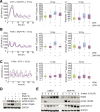
DNA-PKcs inhibition induces an amplified p53 response. (A) Individual A549 cells were tracked, and the average nuclear fluorescence was measured upon 10-Gy γ-IR in cells treated with ATMi, ATRi, or DNA-PKi as indicated. (B–D) Quantification of the relative amplitude and full-width at half-maximum (FWHM) of the first p53 pulse in A549 cells upon (B) 10-Gy γ-IR in cells untreated or treated individually with ATMi, ATRi, DNA-PKi, or DNA-PKi-2 (>130 cells/condition), (C) 2.5, 5, or 10 Gy in cells untreated or treated with DNA-PKi (>730 cells/condition), or (D) 10-Gy γ-IR untreated or treated with LigIV inhibitor (>170 cells/condition). For statistical analysis, see Supplemental Figures S2 and S3.
FIGURE 3:
FIGURE 3:
Loss of DNA-PKcs activity modulates the p53 response through hyperactivation of ATM. (A–C) Individual trajectories and quantification of the relative amplitude and FWHM of the first p53 pulse upon 10-Gy γ-IR in A549 cells untreated or treated with (A) DNA-PKi and ATRi (>280 cells/condition), (B) DNA-PKi and ATMi (>260 cells/condition), or (C) ATMi and ATRi (>260 cells/condition) alone or in combination. (D) Analysis of ATM activity (measured by Western blotting with pATM- and pChk2-specific antibodies) in A549 cells untreated or treated with DNA-PKi and ATMi alone or a combination of both, followed by 10-Gy γ-IR. Asterisk indicates a nonspecific band. (E) Western blot analysis of Mdm2, pChk2, pp53, and GAPDH upon 10-Gy γ-IR in A549 cells untreated or treated with DNA-PKi. For statistical analysis, see Supplemental Figure S3.
FIGURE 4:
FIGURE 4:
Prolonged p53 activation leads to altered cell fate decision upon DSB induction. (A) mRNA expression of p53 target genes p21 and YPEL3 was measured at days 1 and 2 in unirradiated A549 cells or upon 5-Gy γ-IR in cells untreated or treated with DNA-PKi. β-Actin was used as an internal control. Error bars indicate SD of technical triplicates. (B) Percentage of dividing A549 cells during 3 d after damage (5-Gy γ-IR) in wild-type (WT) or p53-knockdown (KD) conditions in the presence or absence of DNA-PKi. Error bars indicate SE of sample proportion. Initial number of cells, >125/condition. (C) Representative images of single A549 cells during 3 d upon 5-Gy γ-IR in cells untreated or treated with DNA-PKi in WT or p53 KD conditions. Blue boxes highlight the senescence-like phenotype observed in p53 WT cells treated with DNA-PKi.

References

    1. Batchelor E, Mock CS, Bhan I, Loewer A, Lahav G. Recurrent initiation: a mechanism for triggering p53 pulses in response to DNA damage. Mol Cell. 2008;30:277–289. - PMC - PubMed
    1. Boehme KA, Kulikov R, Blattner C. p53 stabilization in response to DNA damage requires Akt/PKB and DNA-PK. Proc Natl Acad Sci USA. 2008;105:7785–7790. - PMC - PubMed
    1. Borcherds W, Theillet F-X, Katzer A, Finzel A, Mishall KM, Powell AT, Wu H, Manieri W, Dieterich C, Selenko P, et al. Disorder and residual helicity alter p53-Mdm2 binding affinity and signaling in cells. Nat Chem Biol. 2014;10:1000–1002. - PubMed
    1. Brummelkamp TR, Bernards R, Agami R. Stable suppression of tumorigenicity by virus-mediated RNA interference. Cancer Cell. 2002;2:243–247. - PubMed
    1. Callén E, Jankovic M, Wong N, Zha S, Chen H-T, Difilippantonio S, Di Virgilio M, Heidkamp G, Alt FW, Nussenzweig A, et al. Essential role for DNA-PKcs in DNA double-strand break repair and apoptosis in ATM-deficient lymphocytes. Mol Cell. 2009;34:285–297. - PMC - PubMed

MeSH terms

LinkOut - more resources