. 2016 Aug 1;76(15):4470-80.
doi: 10.1158/0008-5472.CAN-15-2949. Epub 2016 Jun 8.

Eradication of Acute Myeloid Leukemia with FLT3 Ligand-Targeted miR-150 Nanoparticles

Affiliations

Eradication of Acute Myeloid Leukemia with FLT3 Ligand-Targeted miR-150 Nanoparticles

Xi Jiang et al. Cancer Res. .

Abstract

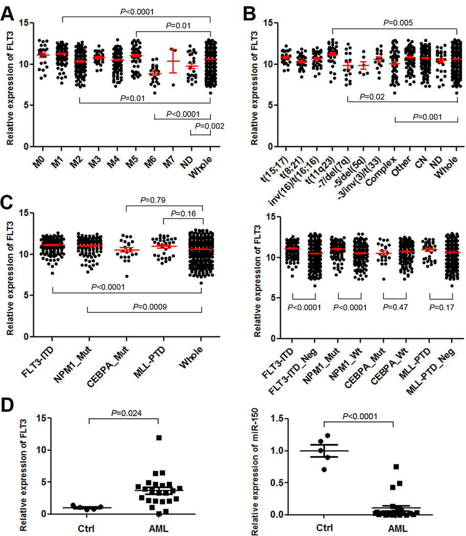
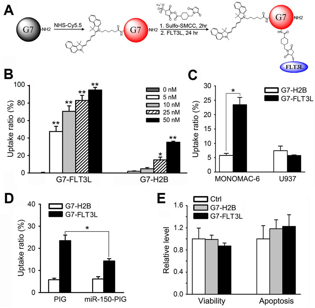
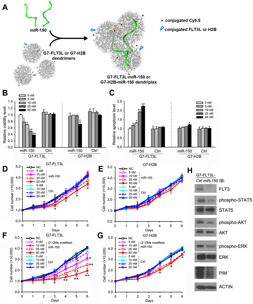
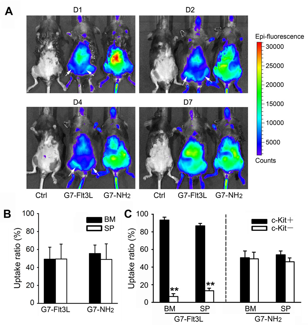
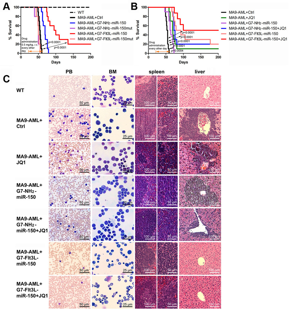
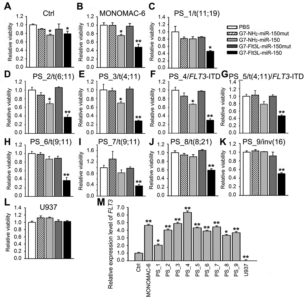
Acute myeloid leukemia (AML) is a common and fatal form of hematopoietic malignancy. Overexpression and/or mutations of FLT3 have been shown to occur in the majority of cases of AML. Our analysis of a large-scale AML patient cohort (N = 562) indicates that FLT3 is particularly highly expressed in some subtypes of AML, such as AML with t(11q23)/MLL-rearrangements or FLT3-ITD. Such AML subtypes are known to be associated with unfavorable prognosis. To treat FLT3-overexpressing AML, we developed a novel targeted nanoparticle system: FLT3 ligand (FLT3L)-conjugated G7 poly(amidoamine) (PAMAM) nanosized dendriplex encapsulating miR-150, a pivotal tumor suppressor and negative regulator of FLT3 We show that the FLT3L-guided miR-150 nanoparticles selectively and efficiently target FLT3-overexpressing AML cells and significantly inhibit viability/growth and promote apoptosis of the AML cells. Our proof-of-concept animal model studies demonstrate that the FLT3L-guided miR-150 nanoparticles tend to concentrate in bone marrow, and significantly inhibit progression of FLT3-overexpressing AML in vivo, while exhibiting no obvious side effects on normal hematopoiesis. Collectively, we have developed a novel targeted therapeutic strategy, using FLT3L-guided miR-150-based nanoparticles, to treat FLT3-overexpressing AML with high efficacy and minimal side effects. Cancer Res; 76(15); 4470-80. ©2016 AACR.


Conflict of interest statement

Confliction of Interest: The authors declare no conflict of interest.

Figures

Figure 1
Figure 1. FLT3 is highly expressed and miR-150 is repressed in AML
(A–C) The expression patterns of FLT3 across different FAB subtypes (A), cytogenetic subtypes (B) or subtypes with different molecular mutations (C) in the GSE37642 AML cohort. In Figure plots A and B, P-value was shown for the particular AML subtypes in which FLT3 was expressed at a significantly higher or lower level than that in the whole AML set (n=562). In Figure plot C, the P-value was shown for comparison between each individual AML subtype (with a particular type of molecular abnormalities) and the whole AML set (left panel), or between AML cases with and without a particular type of molecular abnormalities (right panel). ND, not determined; complex, complex karyotypes; other, other cytogenetic abnormalities; CN, cytogenetically normal; _Mut, with a particular type of molecular mutations; _Neg, without a particular type of molecular mutations. Note: The expression values were log(2) transformed and normalized by RMA (29). Over 40% of the AML cases with NPM1 mutations also have FLT3-ITD. (D) qPCR analysis of expression of both FLT3 and miR-150 in 23 human primary AML mononuclear (MNC) samples including 3 t(4;11), 1 t(6;11), 9 t(9;11), 1 t(11;19), 3 t(8;21), 1 t(15;17), 3 inv(16), 1 FLT3-ITD, and 1 t(4;11)/FLT3-ITD, along with 5 normal BM cell controls (Ctrl; including 3 MNC and 2 CD34+ samples). The P-values were calculated by two-tailed t-test.
Figure 2
Figure 2. Development of G7-FLT3L dendrimers and their selective targeting to FLT3-overexpressing AML cells
(A) G7-NH2 PAMAM dendrimers were first conjugated with the near-infrared dye Cy5.5 using an N-hydroxysuccinimide ester linker sulfo-SMCC. Next, the fluorescently-tagged G7-NH2 (G7-Cy5.5) was conjugated to human recombinant FLT3L (i.e., the soluble FLT3L form with 155 amino acids; ProSpec-Tany Technogene Ltd., East Brunswick, NJ) at a 1:2 ratio using the heterofunctional linker sulfosuccinimidyl 4-((N-maleimidomethyl) cyclohexane-1-carboxylate) (sulfo-SMCC), resulting in the G7-FLT3L conjugates. (B) MONOMAC-6 cells were treated with Cy5.5-conjugated G7-FLT3L or G7-H2B nanoparticles for 24 hrs at the indicated doses. The proportion of Cy5.5+ cells were detected through flow cytometry analysis. (C) MONOMAC-6 and U937 cells were treated with 50 nM Cy5.5-conjugated G7-FLT3L or G7-H2B nanoparticles for 1 hr. The proportion of Cy5.5+ cells were detected through flow cytometry analysis. (D) MONOMAC-6 cells transfected with MSCV-PIG-miR-150 (miR-150-PIG) or MSCV-PIG (PIG) were treated with 50 nM Cy5.5-conjugated G7-FLT3L or G7-H2B nanoparticles for 1 hr. The proportion of Cy5.5+ cells were detected through flow cytometry analysis. Each experiment was repeated independently for at least three times. Average levels of at least three replicates are shown. (E) Cell viability (left panel) and apoptosis (right panel) of MONOMAC-6 cells treated with PBS (Ctrl), 50 nM G7-FLT3L or G7-H2B nanoparticles for 48 hr. *, P<0.05; **, P<0.01.
Figure 3
Figure 3. The formation of the G7-FLT3L-miR-150 nanoparticles and their inhibitory effect on MONOMAC-6 cell growth, as well as the FLT3 signaling pathway in vitro
(A) G7-FLT3L dendrimers were integrated with miR-150 oligos to form stabilized G7-FLT3L-miR-150 dendriplexes. (B, C) MONOMAC-6 cells were treated with G7-FLT3L or G7-H2B nanoparticles complexed with miR-150 or miR-150 mutant RNA (i.e., Control/Ctrl) oligos at the indicated doses. Cell viability (B) or cell apoptosis (C) 48 hrs post treatment are shown. (D, E) The treated cells were counted at indicated time points post-drug treatment. Total cell numbers of G7-FLT3L (D) or G7-H2B (E) treated groups are shown. (F, G) MONOMAC-6 cells were treated with G7-FLT3L or G7-H2B nanoparticles complexed with 2’-OMe-modified miR-150 or miR-150 mutant RNA oligos at the indicated doses. Total cell numbers of G7-FLT3L (F) or G7-H2B (G) treated groups at the indicated time points are shown. *, P<0.05; **, P<0.01. (H) MONOMAC-6 cells were treated with 50 nM G7-FLT3L-miR-150 or G7-FLT3L control (Ctrl) nanoparticles for 72 hours. Levels of FLT3, phosphorylated STAT5 (Y694), STAT5, phosphorylated AKT (S473), AKT, phosphorylated ERK (T202/Y204), ERK, PIM and ACTIN were determined by Western blotting.
Figure 4
Figure 4. In vivo distribution of the G7-Flt3L or G7-NH2
(A) C57BL/6 wild type mice were treated with 0.5 mg/kg Cy5.5-conjugated G7-Flt3L or G7-NH2, i.v., once. Whole animal images at the indicated time points are shown (D1 = day 1, etc.). The white arrows indicate the location of femur bone marrow (BM). (B) Uptake ratio of Cy5.5-conjugated G7-Flt3L or G7- NH2 in mouse BM and spleen (SP) cells. Animals were sacrificed 7 days post treatment, and the Cy5.5+ ratio was determined through flow cytometry analysis. (C) Uptake ratio of Cy5.5-conjugated G7-Flt3L or G7-NH2 in mouse BM and SP c-Kit+ or c-Kit cells. Cy5.5+ ratio in c-Kit+ or c-Kit cell populations is shown. Each experiment was repeated independently for at least three times. Average levels of at least three replicates are shown. *, P<0.05.
Figure 5
Figure 5. Therapeutic effect of G7-Flt3L-miR-150 nanoparticles in treating AML
(A) Secondary BMT recipient mice transplanted with primary MLL-AF9 AML cells were treated with nanoparticles (G7-NH2-miR-150mut, G7-NH2-miR-150, G7-Flt3L-miR-150mut or G7-Flt3L-miR-150) or PBS control after the onset of AML. The medium survivals of G7-Flt3L-miR-150 group, the control group and the G7-NH2-miR-150 treated group are 86 days, 54 days, and 63 days, respectively. (B) Synergistic therapeutic effect of G7-Flt3L-miR-150 and JQ1. The same MLL-AF9 AML mouse model was employed. Kaplan-Meier curves are shown. G7-NH2-miR-150+JQ1 v.s. JQ1 alone: P=0.2062; G7-Flt3L-miR-150 v.s. JQ1 alone: P=0.0051. All the P values were detected by log-rank test. (C) Wright-Giemsa stained PB and BM, and hematoxylin and eosin (H&E) stained spleen and liver of the MLL-AF9-secondary leukemic mice treated with PBS control, JQ1 and/or miR-150-formulated nanoparticles. Samples were taken at the endpoints or 200 days post-BM transplantation. Representative images are shown.
Figure 6
Figure 6. Broad inhibitory effects of G7-Flt3L-miR-150 nanoparticles on AML cell viability
(A–L) Cell viability of BM mononuclear cells from a healthy donor (A), MONOMAC-6 cells (B), BM mononuclear cells from AML patients bearing t(11;19) (C), t(6;11) (D), t(4;11) (E), FLT3-ITD (F), t(4;11)/FLT3-ITD (G), t(9;11) (H and I), t(8;21) (J), inv(16) (K) and U937 cells (L). Cells were treated with PBS, 50 nM G7-NH2-miR-150mut, G7-NH2-miR-150, G7-Flt3L-miR-150mut or G7-Flt3L-miR-150. Cell viability was tested through MTS assays 48 hrs post treatment. (M) Relative gene expression level of FLT3 in the above samples. *, P<0.05; **, P<0.01.

References

    1. Dohner H, Weisdorf DJ, Bloomfield CD. Acute Myeloid Leukemia. N Engl J Med. 2015;373:1136–1152. - PubMed
    1. Lyman SD, James L, Vanden Bos T, de Vries P, Brasel K, Gliniak B, et al. Molecular cloning of a ligand for the flt3/flk-2 tyrosine kinase receptor: a proliferative factor for primitive hematopoietic cells. Cell. 1993;75:1157–1167. - PubMed
    1. Bullinger L, Dohner K, Kranz R, Stirner C, Frohling S, Scholl C, et al. An FLT3 gene-expression signature predicts clinical outcome in normal karyotype AML. Blood. 2008;111:4490–4495. - PubMed
    1. Whitman SP, Maharry K, Radmacher MD, Becker H, Mrozek K, Margeson D, et al. FLT3 internal tandem duplication associates with adverse outcome and gene- and microRNA-expression signatures in patients 60 years of age or older with primary cytogenetically normal acute myeloid leukemia: a Cancer and Leukemia Group B study. Blood. 2010;116:3622–3626. - PMC - PubMed
    1. Kayser S, Levis MJ. FLT3 tyrosine kinase inhibitors in acute myeloid leukemia: clinical implications and limitations. Leuk Lymphoma. 2014;55:243–255. - PMC - PubMed

Publication types