. 2016 Jun 9;14(6):e1002480.
doi: 10.1371/journal.pbio.1002480. eCollection 2016 Jun.

The DEAD-box Protein Rok1 Orchestrates 40S and 60S Ribosome Assembly by Promoting the Release of Rrp5 from Pre-40S Ribosomes to Allow for 60S Maturation

Affiliations

The DEAD-box Protein Rok1 Orchestrates 40S and 60S Ribosome Assembly by Promoting the Release of Rrp5 from Pre-40S Ribosomes to Allow for 60S Maturation

Sohail Khoshnevis et al. PLoS Biol. .

Abstract

DEAD-box proteins are ubiquitous regulators of RNA biology. While commonly dubbed "helicases," their activities also include duplex annealing, adenosine triphosphate (ATP)-dependent RNA binding, and RNA-protein complex remodeling. Rok1, an essential DEAD-box protein, and its cofactor Rrp5 are required for ribosome assembly. Here, we use in vivo and in vitro biochemical analyses to demonstrate that ATP-bound Rok1, but not adenosine diphosphate (ADP)-bound Rok1, stabilizes Rrp5 binding to 40S ribosomes. Interconversion between these two forms by ATP hydrolysis is required for release of Rrp5 from pre-40S ribosomes in vivo, thereby allowing Rrp5 to carry out its role in 60S subunit assembly. Furthermore, our data also strongly suggest that the previously described accumulation of snR30 upon Rok1 inactivation arises because Rrp5 release is blocked and implicate a previously undescribed interaction between Rrp5 and the DEAD-box protein Has1 in mediating snR30 accumulation when Rrp5 release from pre-40S subunits is blocked.


Conflict of interest statement

The authors have declared that no competing interests exist.

Figures

Fig 1
Fig 1. Rrp5 binds to 40S ribosomal subunits in vivo and in vitro.
(A) Ultracentrifugation pelleting experiments demonstrate that recombinant full-length Rrp5 binds purified mature 40S subunits in vitro. S, supernatant; P, pellet. (B) The three C-terminal S1 domains are essential for the interaction between Rrp5 and 40S subunits in vitro. Recombinant Rrp5 fragments are used in the same pelleting experiment as 1A. (C) Gradient centrifugation demonstrates a role for the three C-terminal S1 domains and S1–S5 for binding to preribosomes in vivo. (D) Gradient sedimentation experiments of various recombinant Rrp5•Rok1•AMPPCP complexes demonstrate that S1–S5 contributes to Rrp5 binding to 40S ribosomes in vitro. Note that the more sensitive gradient sedimentation experiments are required to demonstrate the more quantitative than qualitative differences in Rrp8_C8 and Rrp5_C7 binding. Because Rrp5_N9 and Rrp5_N12 do not bind Rok1, binding of these fragments requires the pelleting assay. All experiments were repeated at least twice, and representative data are shown.
Fig 2
Fig 2. Rok1•Rrp5 binds to 40S subunits in the ATP-bound form.
(A) Glycerol density gradient followed by protein precipitation and western blotting was used to analyze the binding of recombinant Rrp5•Rok1 to 40S subunits in the presence of AMPPCP or ADP. Fractions 1 and 13 represent the top (5% glycerol) and the bottom (20% glycerol) of the gradient, respectively. (B) Quantitation of the data in panel A and S2B Fig. The data are averages from two biological replicates. The numerical data underlying this graph can be found in S1 Data. (C) In the absence of 40S ribosomes, neither Rok1 nor Rrp5 enter the gradient, demonstrating that cosedimentation with 40S ribosomes reflects 40S binding. (D) Control gradients in the absence of Rok1 demonstrate that the AMPPCP- and ADP-dependent effects are mediated by Rok1. (E) Rok1 alone does not bind to 40S subunits in the presence of either AMPPCP or ADP. All experiments were repeated at least twice, and representative gels are shown. (F) Electrophoretic mobility shift assay of Rrp5_C7•Rok1 to H44-A2 rRNA mimics in the presence of AMPPCP or ADP. The left panel shows quantitation of data on the right, fit with a single binding isotherm to yield K1/2 values for the AMPPCP and ADP states of 57 ± 7 nM and 106 ± 24 nM, respectively. The numerical data underlying this graph can be found in S1 Data. Error bars come from three independent experiments.
Fig 3
Fig 3. ATP-hydrolysis by Rok1 is required for Rrp5 release from pre-40S ribosomes in vivo.
(A) Western and northern blot analysis of pre-40S ribosomes purified from yeast cells with Enp1-TAP reveals the accumulation of Rrp5, Has1, and snR30 in pre-40S complexes isolated from Rok1 ATPase-deficient cells relative to the WT cells. The TAP-antibody was used as a loading control. (B) Northern blot analysis of preribosomes captured with Rrp5-TAP in the presence of wild-type Rok1 (WT) or ATPase-deficient mutants of Rok1 (K172A and D280A) reveals decreased binding to pre-60S subunits (containing 27S pre-rRNAs) relative to pre-40S subunits (containing 23S pre-rRNA). The data from three biological replicates were quantitated and normalized such that in wild-type cells the 27S/23S levels are set to 1 and the mutants are expressed relative to the WT. The numerical data underlying this graph can be found in S1 Data.
Fig 4
Fig 4. The structure of Rrp5 complexes.
3-D reconstructions of (A) Rrp5•Rok1 or (B) Rrp5•RNA. 90° rotations of Rrp5 complexes around its long axis show the curved, thumb-like projection and hollow fist-like body. The area corresponding to Rok1’s RecA motifs is shaded in blue. By Fourier shell correlation (FSC) = 0.5, the structures are each at about 2.8 nm resolution (S9A Fig). In the final column, the asterisk marks the tip of the TPR.
Fig 5
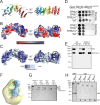
Fig 5. The structure of the TPR domain of Rrp5.
(A) Cartoon representation of the crystal structure. (B) Surface charge distribution of the TPR domain of Rrp5. (C) Surface representation of amino acid conservation calculated using the ConSurf server [35] and displayed in PyMOL (The PyMOL Molecular Graphics System, Version 1.0r1 Schrödinger). The program ConSurf uses the provided sequence (S. cerevisiae Rrp5) to find the 150 closest homologs (lowest E-values) for generating a multiple sequence alignment. The organisms used for Rrp5-TPR can be found here: http://consurf.tau.ac.il/results/1463756501/query_final_homolougs.html. The conserved patches on the surface of the TPR motif that interact with Rok1 are marked in red. (D) Mutations in conserved charged areas of the TPR domain are deleterious in vivo. TPR2: E1509K/E1510K/E1512K and TPR7: K1686E/K1689E. (E) Mutations in conserved charged areas of the TPR domain weaken the Rok1•Rrp5 interaction in vitro. (F) Position of the TPR mutations in the 3-D reconstruction of the Rrp5•Rok1 complex. (G) Recombinant GST-Rok1 immobilized on glutathione (GSH) sepharose resin interacts with recombinant, purified Rrp5FL, Rrp5_C7, and Rrp5_C6, but not Rrp5_C5. (H) Maltose binding protein (MBP) or glutathione S-transferase (GST) alone does not bind to the Rrp5 fragments tested here. I, input; W, wash; E, elution.
Fig 6
Fig 6. Has1 and snR30 accumulate in pre-40S subunits when the Rok1•Rrp5 interaction is disrupted.
(A) Western and northern blot analysis of pre-40S ribosomes purified with Enp1-TAP from yeast cells expressing wild-type Rrp5 or TPR2+7-mutant Rrp5. The TAP-antibody was used as a loading control. (B) Quantitation of two biological replicates of data on the left. The difference in copurification of the named AFs with mutant and wild-type Rrp5 is shown. The numerical data underlying this graph can be found in S1 Data. (C) Recombinant Has1 binds recombinant His6-Rrp5, but not recombinant MBP-Rok1, and recombinant MBP-Has1 does not bind recombinant Rok1. Has1 alone does not bind to the Ni beads.
Fig 7
Fig 7. Speculative model for Rrp5•Rok1 function during 40S ribosome maturation.
Rrp5 (in magenta) is bound to pre-40S subunits (green) and interacts with ITS1 (grey). Rok1 (in yellow) is recruited to pre-40S subunits (I),and then forms the inhibitory duplex to block premature 3′-end formation (II, different secondary structure shown in lighter grey, indicated by asterisk). After cleavage in ITS1 (III, marked by arrow), ATP (T) is hydrolyzed to ADP (D, IV), leading to dissociation of Rrp5•Rok1 from pre-40S subunits (V).

Similar articles

Cited by

References

    1. Woolford JL Jr., Baserga SJ. Ribosome biogenesis in the yeast Saccharomyces cerevisiae. Genetics. 2013;195(3):643–81. Epub 2013/11/06. 10.1534/genetics.113.153197 - DOI - PMC - PubMed
    1. Kos M, Tollervey D. Yeast pre-rRNA processing and modification occur cotranscriptionally. Molecular cell. 2010;37(6):809–20. Epub 2010/03/30. S1097-2765(10)00206-6 [pii] 10.1016/j.molcel.2010.02.024 - DOI - PMC - PubMed
    1. Osheim YN, French SL, Keck KM, Champion EA, Spasov K, Dragon F, et al. Pre-18S ribosomal RNA is structurally compacted into the SSU processome prior to being cleaved from nascent transcripts in Saccharomyces cerevisiae. Mol Cell. 2004;16(6):943–54. . - PubMed
    1. Arcus V. OB-fold domains: a snapshot of the evolution of sequence, structure and function. Curr Opin Struct Biol. 2002;12(6):794–801. Epub 2002/12/31. . - PubMed
    1. Woolls HA, Lamanna AC, Karbstein K. The Roles of Dim2 in Ribosome Assembly. J Biol Chem. 2011;286: 2578–86. 10.1074/jbc.M110.191494 - DOI - PMC - PubMed

MeSH terms

LinkOut - more resources