. 2016 Jun 8;8(342):342ra78.
doi: 10.1126/scitranslmed.aaf3634.

α-Synuclein binds to TOM20 and inhibits mitochondrial protein import in Parkinson's disease

Affiliations

α-Synuclein binds to TOM20 and inhibits mitochondrial protein import in Parkinson's disease

Roberto Di Maio et al. Sci Transl Med. .

Abstract

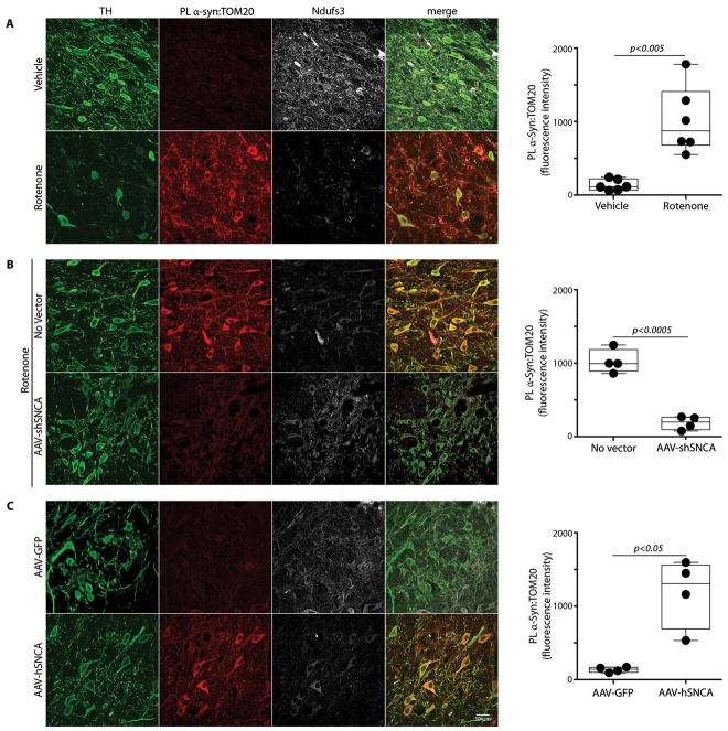
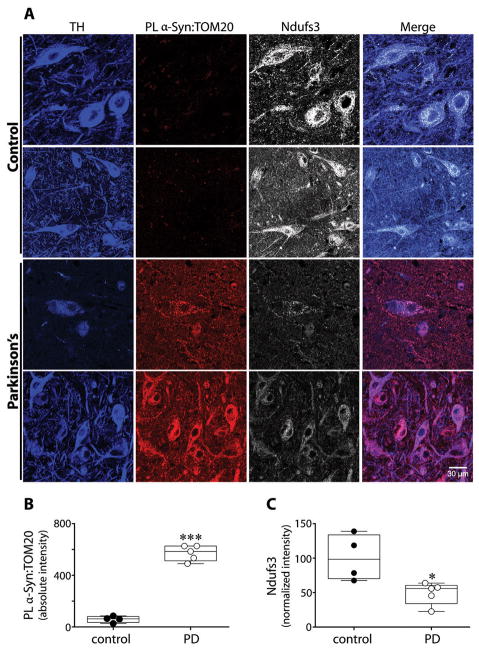
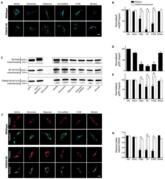
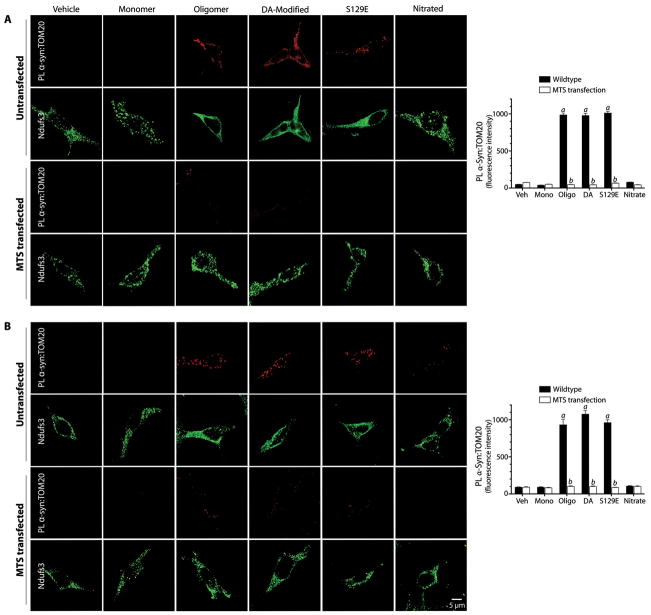
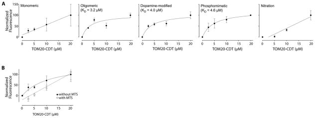
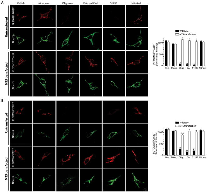
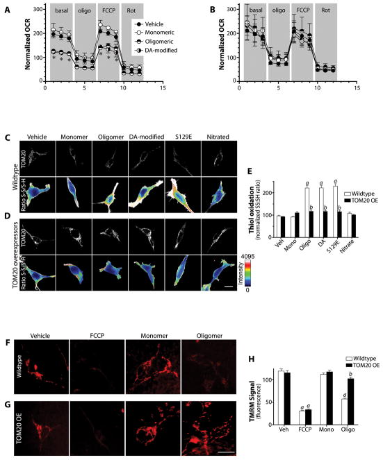
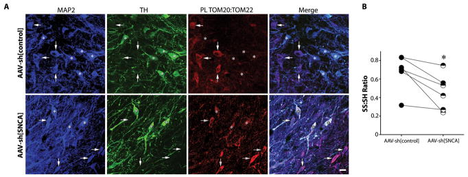
α-Synuclein accumulation and mitochondrial dysfunction have both been strongly implicated in the pathogenesis of Parkinson's disease (PD), and the two appear to be related. Mitochondrial dysfunction leads to accumulation and oligomerization of α-synuclein, and increased levels of α-synuclein cause mitochondrial impairment, but the basis for this bidirectional interaction remains obscure. We now report that certain posttranslationally modified species of α-synuclein bind with high affinity to the TOM20 (translocase of the outer membrane 20) presequence receptor of the mitochondrial protein import machinery. This binding prevented the interaction of TOM20 with its co-receptor, TOM22, and impaired mitochondrial protein import. Consequently, there were deficient mitochondrial respiration, enhanced production of reactive oxygen species, and loss of mitochondrial membrane potential. Examination of postmortem brain tissue from PD patients revealed an aberrant α-synuclein-TOM20 interaction in nigrostriatal dopaminergic neurons that was associated with loss of imported mitochondrial proteins, thereby confirming this pathogenic process in the human disease. Modest knockdown of endogenous α-synuclein was sufficient to maintain mitochondrial protein import in an in vivo model of PD. Furthermore, in in vitro systems, overexpression of TOM20 or a mitochondrial targeting signal peptide had beneficial effects and preserved mitochondrial protein import. This study characterizes a pathogenic mechanism in PD, identifies toxic species of wild-type α-synuclein, and reveals potential new therapeutic strategies for neuroprotection.


Conflict of interest statement

The authors declare that they have no competing financial interests.

Figures

Figure 1
Figure 1
Ex vivo proximity ligation between α-synuclein and TOM20 is associated with decreased mitochondrial import of the complex I subunit, Ndufs3, in nigrostriatal neurons in vivo in the rotenone and α-synuclein overexpression models of PD. (A) In a vehicle-treated rat (top row), there is little α-synuclein:TOM20 PL signal and there is intense punctate staining of Ndufs3 in mitochondria of nigrostriatal neurons. In contrast, in a rotenone-treated rat (bottom row), there is a strong α-synuclein:TOM20 PL signal, which is associated with loss of mitochondrial Ndufs3 staining. In the box plot, mean nigrostriatal cellular PL fluorescence values for individual animals (vehicle- or rotenone-treated) are indicated by black circles. In each animal, PL signal was measured in 35–50 nigrostriatal neurons per hemisphere. Statistical testing by 2-tailed unpaired t-test with Welch’s correction. (B) In a rat that received a unilateral injection of AAV-shSNCA, the rotenone-induced α-synuclein:TOM20 PL signal was largely prevented, and mitochondrial Ndufs3 staining was preserved. In the box plot, mean nigrostriatal cellular PL fluorescence values for each hemisphere (control or AAV-shSNCA-injected) of individual animals are indicated by black circles. For each animal, PL signal was measured in 50–70 nigrostriatal neurons per hemisphere. Statistical testing by 2-tailed paired t-test with Welch’s correction. (C) In a rat that received unilateral injection of an α-synuclein overexpression vector (AAV-hSNCA), the α-synuclein-injected hemisphere shows a strong α-synuclein:TOM20 PL signal with an associated loss of Ndufs3 staining. In the box plot, mean nigrostriatal cellular PL fluorescence values for each hemisphere (control AAV-GFP or AAV-SNCA-injected) of individual animals are indicated by black circles. For each animal, PL signal was measured in 50–70 nigrostriatal neurons per hemisphere. Statistical testing by 2-tailed unpaired t-test with Welch’s correction. TH, tyrosine hydroxylase. Scale bar = 30 μm.
Figure 2
Figure 2
Evidence of impaired mitochondrial protein import in human dopaminergic substantia nigra neurons in Parkinson’s disease. (A) In TH+ dopamine neurons from PD cases, there was an intense α-synuclein:TOM20 PL signal and a marked loss of Ndufs3 immunoreactivity. In PD cases, remaining Ndufs3 staining was rather diffuse instead of punctate. (B) Quantification of the α-synuclein:TOM20 PL signal in control vs. PD dopamine neurons. (C) Quantification of Ndufs3 immunoreactivity in control vs. PD dopamine neurons. The Ndufs3 signal was normalized to the TH signal, which tends to minimize the apparent differences. ***p<0.0001; *p <0.05; 2-tailed unpaired t-test with Welch’s correction for unequal variances.
Figure 3
Figure 3
Post-translationally-modified α-synuclein binds to TOM20 and inhibits mitochondrial protein import. (A) mtGFP import in intact wildtype and TOM20 overexpressing (OE) SH-SY5Y cells exposed to various forms of α-synuclein. In wildtype cells treated with oligomeric, dopamine-modified or S129E α-synuclein, note the diffuse pattern of staining compared to vehicle. In TOM20 overexpressing cells, mtGFP maintained its mitochondrial localization despite α-synuclein treatment. (B) Quantification of mtGFP import in wildtype and TOM20 OE cells. For each condition in each experiment, mtGFP localization was determined in 5–10 ROIs in zoomed confocal images from 5–10 cells, and 3 or 4 independent experiments were performed. (C) Autoradiographs of in vitro import of pre-OTC into mitochondria isolated from rat brain (top), wildtype (WT) SH-SY5Y cells (middle), and TOM20 OE SH-SY5Y cells (bottom) after exposure to various forms of α-synuclein (30 min @ 4°C). Upper band represents 35S-labeled pre-OTC and lower band represents imported, cleaved (mature) OTC. (D) Quantification of OTC import into brain mitochondria. Results were normalized to the vehicle-treated control and FCCP + oligomycin was used to collapse membrane potential and define zero import. N=3 independent experiments. (E) Quantification of OTC import into mitochondria from wildtype and TOM20 overexpressing SH-SY5Y cells. N=3–4 independent experiments. (F) Immunolocalization of TOM20 and Ndufs3 in wildtype and TOM20 overexpressing HEK293 cells exposed to various forms of α-synuclein. In wildtype cells exposed to oligomeric, dopamine-modified or S129E α-synuclein, Ndufs3 localization is diffuse rather than mitochondrial (i.e., Ndufs3 redistributed outside of mitochondria as defined by TOM20). This effect is prevented in TOM20 overexpressing cells. (G) Correlation (Pearson Index) of the localizations of TOM20 and Ndufs3 in wildtype vs. TOM20 overexpressing cells. TOM20 overexpression rescues the normal localization of Ndufs3. For each experimental condition, at least 100 cells were analyzed in each of 3 or 4 independent experiments. Statistical analyses were by by 1- or 2-way ANOVA followed by pairwise testing and correction for multiple comparisons. a – p<0.0001 vs. monomer; b – p<0.0001 vs. wildtype cells; c – p<0.005 vs. monomer; d – p<0.05 vs. wildtype cells; e – p<0.002 vs. wildtype cells. Scale bars = 5 μm.
Figure 4
Figure 4
Proximity ligation of post-translationally modified α-synuclein and TOM20 and Ndufs3 localization in HEK293 cells. (A) In untransfected cells, there is proximity ligation between TOM20 and oligomeric, dopamine-modified and S129E α-synuclein, but not monomeric or nitrated species. This is associated with a cytosolic redistribution of Ndufs3. Fibrillar α-synuclein did not interact with TOM20 (figure S5). When cells were transfected with an MTS expression vector prior to treatment with α-synuclein, the TOM20:α-synuclein interaction was blocked, indicating that the α-synuclein binding site overlaps the MTS binding site on TOM20. MTS transfection also preserved the punctate (mitochondrial) distribution of Ndufs3. (B) When cells were transfected with the MTS expression vector 24h after α-synuclein treatment, the TOM20:α-synuclein interaction was reversed. Bar graphs show quantification of the α-synuclein:TOM20 PL signal in mock transfected (black bars) and MTS-overexpressing cells. At least 100 cells were analyzed for each condition in every independent experiment (N=3). a, p<0.0001 vs vehicle; b, p<0.0001 vs mock transfected; 2-way ANOVA. Scale bar = 5 μm.
Figure 5
Figure 5
Binding curves of TOM20 to various forms of α-synuclein. (A) There was saturable binding of oligomeric, dopamine-modified and S129E α-synuclein, but not the monomeric or nitrated species. N = 3. (B) The COX8 MTS peptide inhibits binding of oligomeric α-synuclein to TOM20. When binding is performed in the presence of excess MTS (250 μM), specific binding is markedly reduced or abolished; nonlinear curve fitting yielded an affinity of >7 × 10¹⁴ μM when the MTS was present. Similar results were obtained with dopamine-modified and S129E α-synuclein. The overall effect of the MTS was significant (p<0.02) by 2-way ANOVA. N=3.
Figure 6
Figure 6
α-Synuclein interaction with TOM20 prevents the normal interaction between TOM20 and TOM22 in HEK293 cells. (A) Under basal conditions (vehicle), proximity ligation detects an interaction between TOM20 and TOM22. This is blocked by oligomeric, dopamine-modified and S129E α-synuclein, but not monomeric or nitrated species. Loss of the TOM20:TOM22 PL signal is associated with relocalization of Ndufs3 to the cytosol. In cells overexpressing a ‘naked’ MTS (COX8 presequence), the TOM20:TOM22 PL signal is maintained even after treatment with oligomeric, dopamine-modified and S129E α-synuclein. (B) When cells were treated with α-synuclein and then transfected with the MTS 24 h later, the TOM20:TOM22 PL signal could be restored. Graphs show quantification of TOM20:TOM22 signal in mock transfected (black bars) and MTS-overexpressing cells (white bars). a, p<0.0001 vs vehicle; b, p<0.0001 vs mock transfected; 2-way ANOVA. At least 100 cells were analyzed per condition in each independent experiment. N=3. Scale bar = 5 μm.
Figure 7
Figure 7
Downstream effects of α-synuclein on mitochondria. (A) In wildtype SH-SY5Y cells, a 24 h exposure to oligomeric or dopamine-modified α-synuclein reduced basal and FCCP-stimulated mitochondrial respiration. Monomeric α-synuclein was without effect. oligo – oligomycin, FCCP - carbonyl cyanide-4-(trifluoromethoxy) phenylhydrazone, Rot – rotenone. *p<0.01 vs vehicle; 1-way ANOVA; N=3. (B) In SH-SY5Y cells overexpressing TOM20, the deleterious effects of oligomeric and dopamine-modified α-synuclein were prevented. N=3. (C) In wildtype HEK293cells, a 24 h exposure to oligomeric, dopamine-modified or S129E α-synuclein induced oxidation of protein thiols; exposure to monomeric or nitrated α-synuclein did not. (D, E) In HEK293 cells overexpressing TOM20, α-synuclein did not induce oxidative stress. At least 100 cells were quantified per condition in each experiment. a, p<0.001 vs vehicle; b, p<0.001 vs mock transfected cells; 2-way ANOVA; N=3. (F) TMRM fluorescence in SH-SY5Y cells (as an index of mitochondrial membrane potential) is reduced by oligomeric but not monomeric α-synuclein. (G, H) In SH-SY5Y cells overexpressing TOM20, oligomeric α-synuclein does not significantly impact mitochondrial membrane potential. 30–50 cells were analyzed for each treatment in each of 3 independent experiments. a – p<0.001 vs. vehicle; b – p< 0.001 vs. wildtype cells; 2-way ANOVA. Scale bars = 10 μm.
Figure 8
Figure 8
The normal TOM20:TOM22 PL signal seen in most neurons is absent in nigrostriatal dopamine neurons in vivo, but is restored by knockdown of endogenous α-synuclein. (A) In the untreated hemisphere (top row), MAP2+/TH- non-dopaminergic neurons (arrows) show a strong TOM20:TOM22 PL signal, which is absent in TH+ dopaminergic cells (asterisks). In the hemisphere that received AAV2-shSNCA (bottom row), there was emergence of a strong TOM20:TOM22 PL signal in the TH+ dopaminergic neurons. Scale bar = 30 μm. (B) Consistent with in vitro data (figure 5C–E), α-synuclein knockdown was associated with decreased levels of protein thiol oxidation. Filled circles, -S-S-/-SH ratio of nigral neurons in the control hemisphere; half-filled circles, -S-S-/-SH ratio of nigral neurons in the SNCA knockdown hemisphere; lines connect the means from each hemisphere in each animal. *p<0.05, Wilcoxon matched-pairs signed rank test. Scale bar = 30 μm.

Comment in

References

    1. Kalia LV, Lang AE. Parkinson’s disease. Lancet. 2015 Apr 17; - PubMed
    1. Zaltieri M, et al. Mitochondrial Dysfunction and alpha-Synuclein Synaptic Pathology in Parkinson’s Disease: Who’s on First? Parkinsons Dis. 2015;2015:108029. - PMC - PubMed
    1. Betarbet R, et al. Chronic systemic pesticide exposure reproduces features of Parkinson’s disease. Nat Neurosci. 2000 Dec;3:1301. - PubMed
    1. Betarbet R, et al. Intersecting pathways to neurodegeneration in Parkinson’s disease: effects of the pesticide rotenone on DJ-1, alpha-synuclein, and the ubiquitin-proteasome system. Neurobiol Dis. 2006 May;22:404. - PubMed
    1. Cannon JR, et al. A highly reproducible rotenone model of Parkinson’s disease. Neurobiol Dis. 2009 May;34:279. - PMC - PubMed

MeSH terms

Substances