. 2016 Jun 8;8(342):342ra79.
doi: 10.1126/scitranslmed.aad8166.

A universal system to select gene-modified hepatocytes in vivo

Affiliations

A universal system to select gene-modified hepatocytes in vivo

Sean Nygaard et al. Sci Transl Med. .

Abstract

Many genetic and acquired liver disorders are amenable to gene and/or cell therapy. However, the efficiencies of cell engraftment and stable genetic modification are low and often subtherapeutic. In particular, targeted gene modifications from homologous recombination are rare events. These obstacles could be overcome if hepatocytes that have undergone genetic modification were to be selectively amplified or expanded. We describe a universally applicable system for in vivo selection and expansion of gene-modified hepatocytes in any genetic background. In this system, the therapeutic transgene is coexpressed with a short hairpin RNA (shRNA) that confers modified hepatocytes with resistance to drug-induced toxicity. An shRNA against the tyrosine catabolic enzyme 4-OH-phenylpyruvate dioxygenase protected hepatocytes from 4-[(2-carboxyethyl)-hydroxyphosphinyl]-3-oxobutyrate, a small-molecule inhibitor of fumarylacetoacetate hydrolase. To select for specific gene targeting events, the protective shRNA was embedded in a microRNA and inserted into a recombinant adeno-associated viral vector designed to integrate site-specifically into the highly active albumin locus. After selection of the gene-targeted cells, transgene expression increased 10- to 1000-fold, reaching supraphysiological levels of human factor 9 protein (50,000 ng/ml) in mice. This drug resistance system can be used to achieve therapeutically relevant transgene levels in hepatocytes in any setting.


Figures

Fig. 1
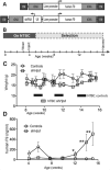
Fig. 1. Identification of an shRNA that rescues Fah deficiency
(A) The tyrosine catabolic pathway. Genetic deficiency of Fah causes hereditary tyrosinemia type 1 (HT1) due to accumulation of fumarylacetoacetate (FAA) in hepatocytes. The disease can be treated pharmacologically (NTBC) or by shRNA knock-down of the genes required for making FAA. CEHPOBA inhibits FAH and causes accumulation of FAA. TAT, tyrosine amino transferase; HPD, 4-OH-phenylpyruvate dioxygenase; HGD, homogentisic acid dioxygenase; MAI, maleylacetoacetate isomerase; FAH, fumarylacetoacetate hydrolase. (B) Lentiviral construct. shRNAs targeting Hpd, Hgd, or Tat were expressed from a U6 promoter. Vector expresses a GFP reporter. (C) Experimental time line. Neonatal Fah-/- mice were injected with shTat, shHpd, shHgd lentiviruses or a vector devoid of any shRNA (control) and kept on NTBC until weaning. NTBC was then withdrawn to permit liver injury and selection of resistant hepatocytes. (D) Mouse weights during selection starting at age 5 weeks. Grey bars represent periods of intermittent NTBC thxrapy. Only the control, shHgd and shTat cohorts were given NTBC after week 6. Data are mean –SD (downward tick) (n=4-6). (E) Mice were injected with a non-selectable vector (control) or shHpd. Liver tissues were stained for the reporter GFP and for α-fetoprotein (AFP), which is highly expressed in mutant Fah-/- hepatocytes. Yellow arrow denotes the absence of AFP within a selected nodule, compared with AFP-positive surrounding tissue (black arrows). Scale bar = 100 μm. (F) PCR amplification of genomic liver DNA with primers flanking the lentiviral-shRNA sequence (two primer sets).
Fig. 2
Fig. 2. Selection of integrating rAAV vectors
(A) Ribosomal rAAV constructs capable of chromosomal integration. (Top) Standard vector control; (bottom) selectable Hpd shRNA vector. Both vectors contain ribosomal DNA (rDNA) homology arms to enhance chromosomal integration, and both expressed human F9. (B) Experimental time line. Selection: Periods of NTBC withdrawal were different between cohorts and are indicated in panel C. (C) Weights and NTBC administration in animals receiving control or shHpd vector. Black rectangles indicate periods of NTBC treatment in control mice, whereas grey bar is NTBC for mice receiving shHpd. Data are means +/- SD (n = 3-5). (D) Plasma human F9 measured by ELISA. Data are means +/- SD (n=4-5). *p<0.05, **p=0.01 versus controls. Differences were evaluated by student two-tailed t-test assuming equal variance.
Fig. 3
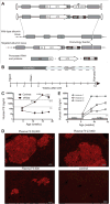
Fig. 3. Selection of targeted integrations in the albumin locus
(A) Generide rAAV constructs designed for chromosomal integration. (Top) The selectable shHpd construct is driven by the pol3 U6 promoter. (Below) The shHpd is embedded within a miRNA and controlled by the endogenous albumin promoter. Both albumin-targeted “generide” vectors encoded human F9 cDNA flanked by mouse albumin homology arms. The structures of the wild-type and gene-targeted albumin locus are also shown. Homologous recombination led to generation of a fused mRNA transcript. RNA processing liberated the shHpd and ribosome skipping at the 2A-peptide coding sequence generated separate mouse albumin (Alb) and hF9 proteins. (B) Experimental time line. Selection cycles starting week 5: off NTBC for 3 weeks, then on for 5 days, until week 20. (C) Plasma human F9 measured by ELISA in mice treated with the selectable or control gene-ride rAAV. The 5% and 100% levels of normal human F9 blood levels are shown with a dashed line. Data are means +/- SD (n = 4). **p<0.01, ***p<0.001 versus controls. Differences were evaluated by student two-tailed t-test assuming equal variance. (D) Human F9 liver immunohistochemistry showing representative nodules from mice with plasma F9 levels of 38,000 and 27,000 ng/ml (high), 800 ng/ml (low), and a control (no selection). Scale bars, 100 μm. (E) Plasma human F9 levels in mice treated with the selectable gene-ride rAAV and subjected to NTBC withdrawal from age 6-9 weeks, followed by reintroduction of NTBC thereafter. The dashed line indicates the therapeutic level of hF9 (250 ng/ml, or 5%). Data are single measurements from individual mice (n = 3).
Fig. 4
Fig. 4. Histology of shHpd selected hF9-positive nodules
(A) Serial sections of representative fields from three separate Fah-/- mice after multiple cycles of selection (NTBC withdrawal). Mice were given either the shHpd vector or saline control. The adjacent sections were stained for expression of hF9, glutamine synthase, a marker for hepatocytes adjacent to the central vein of the hepatic lobule (zone 3), H&E and the proliferation marker Ki67. The “x” and “o” symbols mark vessels as landmarks for lining up the serial sections. For shHpd mouse 1 (shHpd1), serial sections are the same area in low magnification (62×);. For shHpd mouse 2 (shHpd2) and the control mouse, serial sections are high magnification (150×). Black arrows indicate nodules expressing human F9. Scale bars, 100 μm. (B) In vivo selection after gene transfer in adults. Four adult male Fah-/- mice treated from birth with NTBC were injected with 8 × 10¹¹ vg of the generide vector (Fig. 3A) at day 52. NTBC therapy was stopped 10 days later. Human F9 levels were measured. Data are averages +/- SD (n = 4). Differences between hF9 levels at different time points were evaluated by student two-tailed t-test assuming equal variance.
Fig. 5
Fig. 5. Selection of gene-targeted hepatocytes in wild-type mice using pharmacologic FAH inhibition
(A) Experimental time line. Beginning at 4 weeks of age, the mice were given daily i.p. injections of CEHPOBA (1 μmol/g) or saline until 8-weeks of age. (B) Plasma human F9 measured by ELISA in mice treated with CEHPOBA (red symbols) or saline (blue symbols). Data for individual mice are shown. Dashed line denotes 5% of normal F9 levels, which is considered therapeutic. The grey rectangle indicates CEHPOBA treatment periods. A second period of CEHPOBA treatment was administered to only one mouse (inverted triangle). (C) Human F9 immunohistochemistry showing representative liver nodules from two separate CEHPOBA-treated mice and two saline injected controls. Arrows denote hF9-positive hepatocytes. Scale bars, 100 μm. (D) H&E staining of liver and kidney tissues from a representative CEHPOBA-treated animal and saline-treated control. Scale bars, 100 μm.
Fig. 6
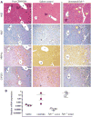
Fig. 6. Histology of CEHPOBA-treated livers
(A) Liver histology of a representative CEHPOBA-treated and a saline control one month after stopping the drug. An untreated age-matched Fah-/- off NTBC (a positive control from a separate experiment) is shown at right for comparison. Serial sections were stained with H&E, followed by antibody stains for Ki67, HNF4α, and CYPE1. Black arrows show portal bile ducts. The “*” marks central veins and “o” delineates portal veins. Pleiocytosis is marked by yellow arrows. (B) Relative abundance of fused transcripts/mouse albumin transcripts from saline-treated C57Bl6 mice, CEHPOBA-treated C57Bl6 mice (8 weeks after completing selection), and NTBC-cycled Fah-/- mice injected with either control vector or the generide vector harboring the shHpd selection cassette (age 20 weeks, see Fig.3C). Data are individual animals (n=3) with means +/- SD.

References

    1. Mingozzi F, High KA. Therapeutic in vivo gene transfer for genetic disease using AAV: progress and challenges. Nat Rev Genet. 2011;12:341–355. - PubMed
    1. Nathwani AC, Tuddenham EG, Rangarajan S, Rosales C, McIntosh J, Linch DC, Chowdary P, Riddell A, Pie AJ, Harrington C, O'Beirne J, Smith K, Pasi J, Glader B, Rustagi P, Ng CY, Kay MA, Zhou J, Spence Y, Morton CL, Allay J, Coleman J, Sleep S, Cunningham JM, Srivastava D, Basner-Tschakarjan E, Mingozzi F, High KA, Gray JT, Reiss UM, Nienhuis AW, Davidoff AM. Adenovirus-associated virus vector-mediated gene transfer in hemophilia B. N Engl J Med. 2011;365:2357–2365. - PMC - PubMed
    1. Wang L, Bell P, Lin J, Calcedo R, Tarantal AF, Wilson JM. AAV8-mediated hepatic gene transfer in infant rhesus monkeys (Macaca mulatta) Mol Ther. 2011;19:2012–2020. - PMC - PubMed
    1. Pien GC, Basner-Tschakarjan E, Hui DJ, Mentlik AN, Finn JD, Hasbrouck NC, Zhou S, Murphy SL, Maus MV, Mingozzi F, Orange JS, High KA. Capsid antigen presentation flags human hepatocytes for destruction after transduction by adeno-associated viral vectors. J Clin Invest. 2009;119:1688–1695. - PMC - PubMed
    1. Mingozzi F, High KA. Immune responses to AAV in clinical trials. Curr Gene Ther. 2007;7:316–324. - PubMed

Publication types