

Anti-EBOV GP IgGs Lacking α1-3-Galactose and Neu5Gc Prolong Survival and Decrease Blood Viral Load in EBOV-Infected Guinea Pigs
- PMID: 27280712
- PMCID: PMC4900587
- DOI: 10.1371/journal.pone.0156775
Anti-EBOV GP IgGs Lacking α1-3-Galactose and Neu5Gc Prolong Survival and Decrease Blood Viral Load in EBOV-Infected Guinea Pigs
Abstract
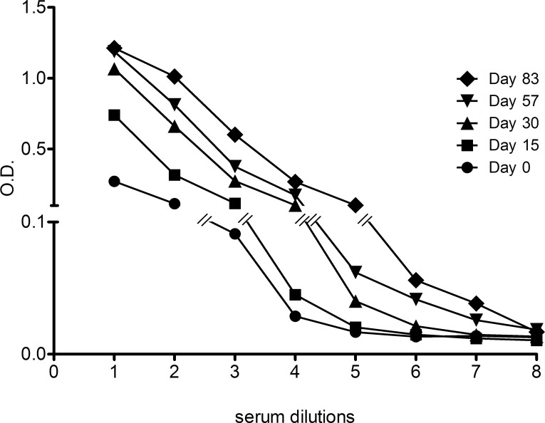
Polyclonal xenogenic IgGs, although having been used in the prevention and cure of severe infectious diseases, are highly immunogenic, which may restrict their usage in new applications such as Ebola hemorrhagic fever. IgG glycans display powerful xenogeneic antigens in humans, for example α1-3 Galactose and the glycolyl form of neuraminic acid Neu5Gc, and IgGs deprived of these key sugar epitopes may represent an advantage for passive immunotherapy. In this paper, we explored whether low immunogenicity IgGs had a protective effect on a guinea pig model of Ebola virus (EBOV) infection. For this purpose, a double knock-out pig lacking α1-3 Galactose and Neu5Gc was immunized against virus-like particles displaying surface EBOV glycoprotein GP. Following purification from serum, hyper-immune polyclonal IgGs were obtained, exhibiting an anti-EBOV GP titer of 1:100,000 and a virus neutralizing titer of 1:100. Guinea pigs were injected intramuscularly with purified IgGs on day 0 and day 3 post-EBOV infection. Compared to control animals treated with IgGs from non-immunized double KO pigs, the anti-EBOV IgGs-treated animals exhibited a significantly prolonged survival and a decreased virus load in blood on day 3. The data obtained indicated that IgGs lacking α1-3 Galactose and Neu5Gc, two highly immunogenic epitopes in humans, have a protective effect upon EBOV infection.
Conflict of interest statement
Figures
Similar articles
-
Ebolavirus Glycoprotein Fc Fusion Protein Protects Guinea Pigs against Lethal Challenge.PLoS One. 2016 Sep 13;11(9):e0162446. doi: 10.1371/journal.pone.0162446. eCollection 2016. PLoS One. 2016. PMID: 27622456 Free PMC article.
-
Distinct Immunogenicity and Efficacy of Poxvirus-Based Vaccine Candidates against Ebola Virus Expressing GP and VP40 Proteins.J Virol. 2018 May 14;92(11):e00363-18. doi: 10.1128/JVI.00363-18. Print 2018 Jun 1. J Virol. 2018. PMID: 29514907 Free PMC article.
-
Matrix-M adjuvant enhances antibody, cellular and protective immune responses of a Zaire Ebola/Makona virus glycoprotein (GP) nanoparticle vaccine in mice.Vaccine. 2016 Apr 7;34(16):1927-35. doi: 10.1016/j.vaccine.2016.02.033. Epub 2016 Feb 24. Vaccine. 2016. PMID: 26921779
-
Therapeutic vaccination strategies against EBOV by rVSV-EBOV-GP: the role of innate immunity.Curr Opin Virol. 2021 Dec;51:179-189. doi: 10.1016/j.coviro.2021.10.007. Epub 2021 Nov 5. Curr Opin Virol. 2021. PMID: 34749265 Free PMC article. Review.
-
The Ebola virus glycoprotein and its immune responses across multiple vaccine platforms.Expert Rev Vaccines. 2020 Mar;19(3):267-277. doi: 10.1080/14760584.2020.1738225. Epub 2020 Mar 16. Expert Rev Vaccines. 2020. PMID: 32129120 Review.
Cited by
-
N-Glycolylneuraminic Acid (Neu5Gc) Null Large Animals by Targeting the CMP-Neu5Gc Hydroxylase (CMAH).Front Immunol. 2019 Oct 15;10:2396. doi: 10.3389/fimmu.2019.02396. eCollection 2019. Front Immunol. 2019. PMID: 31681287 Free PMC article. Review.
-
Successful post-exposure prophylaxis of Ebola infected non-human primates using Ebola glycoprotein-specific equine IgG.Sci Rep. 2017 Feb 3;7:41537. doi: 10.1038/srep41537. Sci Rep. 2017. PMID: 28155869 Free PMC article.
-
High neutralizing potency of swine glyco-humanized polyclonal antibodies against SARS-CoV-2.Eur J Immunol. 2021 Jun;51(6):1412-1422. doi: 10.1002/eji.202049072. Epub 2021 Mar 2. Eur J Immunol. 2021. PMID: 33576494 Free PMC article.
-
The Effects of an In Vitro Oocyte Maturation System and Chlorogenic Acid Supplementation during Embryo Culture on the Development of Porcine Cloned Embryos Derived from Native Vietnamese Ban Pigs.Vet Med Int. 2023 Apr 17;2023:5702970. doi: 10.1155/2023/5702970. eCollection 2023. Vet Med Int. 2023. PMID: 37101560 Free PMC article.
References
-
- Buelow R, van Schooten W. The future of antibody therapy. Ernst Schering Found Symp Proc. 2006; 83–106. - PubMed
MeSH terms
Substances
LinkOut - more resources
Full Text Sources
Other Literature Sources
Medical
Research Materials
