. 2016 Jun 9;11(6):e0157280.
doi: 10.1371/journal.pone.0157280. eCollection 2016.

Extratumoral Heme Oxygenase-1 (HO-1) Expressing Macrophages Likely Promote Primary and Metastatic Prostate Tumor Growth

Affiliations

Extratumoral Heme Oxygenase-1 (HO-1) Expressing Macrophages Likely Promote Primary and Metastatic Prostate Tumor Growth

Sofia Halin Bergström et al. PLoS One. .

Abstract

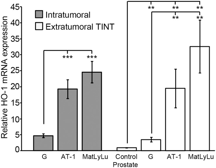
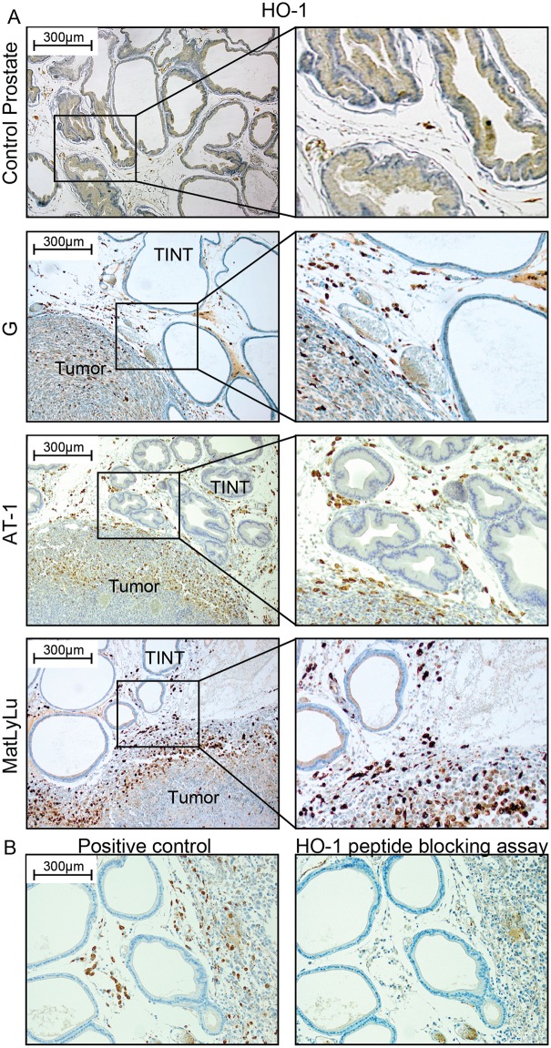
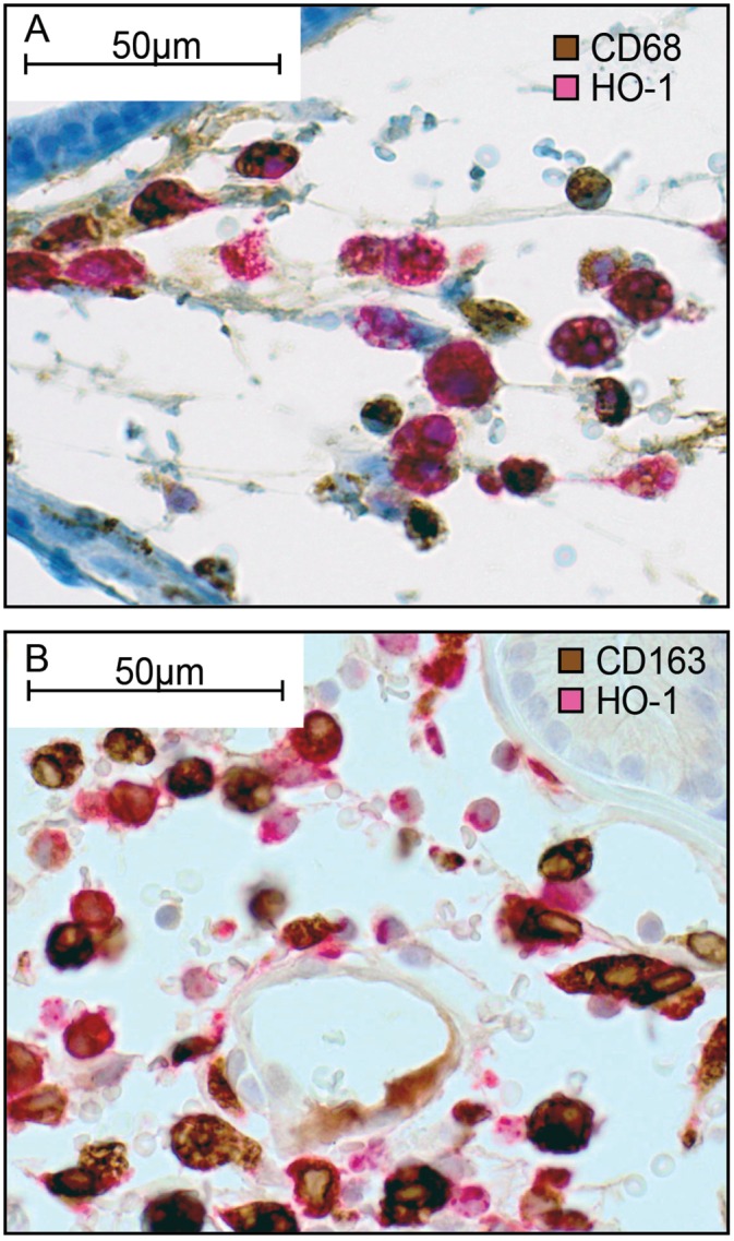
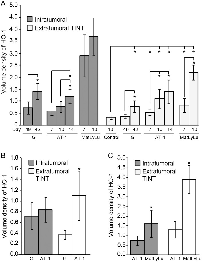
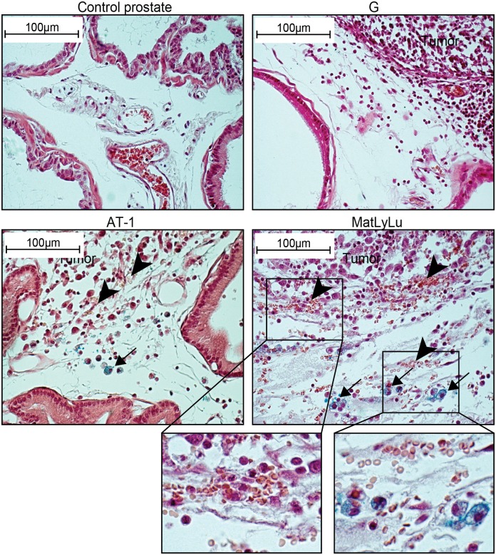
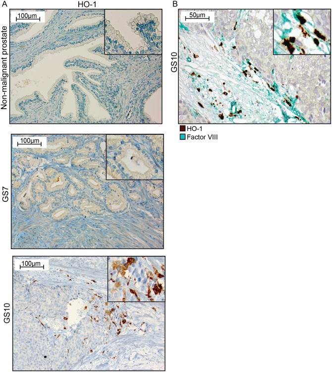
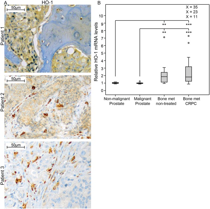
Aggressive tumors induce tumor-supporting changes in the benign parts of the prostate. One factor that has increased expression outside prostate tumors is hemoxygenase-1 (HO-1). To investigate HO-1 expression in more detail, we analyzed samples of tumor tissue and peritumoral normal prostate tissue from rats carrying cancers with different metastatic capacity, and human prostate cancer tissue samples from primary tumors and bone metastases. In rat prostate tumor samples, immunohistochemistry and quantitative RT-PCR showed that the main site of HO-1 synthesis was HO-1+ macrophages that accumulated in the tumor-bearing organ, and at the tumor-invasive front. Small metastatic tumors were considerably more effective in attracting HO-1+ macrophages than larger non-metastatic ones. In clinical samples, accumulation of HO-1+ macrophages was seen at the tumor invasive front, almost exclusively in high-grade tumors, and it correlated with the presence of bone metastases. HO-1+ macrophages, located at the tumor invasive front, were more abundant in bone metastases than in primary tumors. HO-1 expression in bone metastases was variable, and positively correlated with the expression of macrophage markers but negatively correlated with androgen receptor expression, suggesting that elevated HO-1 could be a marker for a subgroup of bone metastases. Together with another recent observation showing that selective knockout of HO-1 in macrophages reduced prostate tumor growth and metastatic capacity in animals, the results of this study suggest that extratumoral HO-1+ macrophages may have an important role in prostate cancer.


Conflict of interest statement

Competing Interests: The authors have declared that no competing interests exist.

Figures

Fig 1
Fig 1. HO-1 mRNA expression in rat prostate tumors and in the surrounding non-malignant prostate tissue (TINT).
HO-1 mRNA expression in rat tumors and TINT expressed in relation to the levels in tumor-free control prostate tissue (**p <0.01, ***p < 0.001, n = 7–8 in each group).
Fig 2
Fig 2. HO-1 protein expression in rat prostate tumors and in the surrounding non-malignant prostate tissue (TINT).
(A) Representative sections of control rat prostate tissue and orthotopic rat prostate tumors and TINT stained for HO-1 (brown) (left panel; 100x magnifications, right panel; shows higher magnifications). (B) Antibody specificity control. No staining was seen in sections incubated with primary antibody that had been pre-incubated with an excess of a recombinant rat HO-1 peptide.
Fig 3
Fig 3. HO-1 expression in macrophages.
Representative sections (400x magnifications) of rat prostate MatLyLu TINT double stained for CD68+ (brow) and HO-1+ (red) cells (A), or double stained for CD163+ (brown) and HO-1+ (red) cells (B). Most cells stained both red and blown suggesting that most HO-1+ cells are macrophages, and in particular of the M2-type (CD163+).
Fig 4
Fig 4. Volume density of HO-1+ cells in rat prostate tumors and in the surrounding non-malignant prostate tissue (TINT).
(A) Volume density of HO-1+ cells–at different time points and tumor sizes (mean tumor weight mg +/- SD)–in G tumors (day 49 (small); 49 +/- 21 mg and day 42 (large); 250 +/- 164 mg), in AT-1 tumors (day 7; 15 +/- 4.5 mg, day 10; 71 +/- 48 mg, and day 14; 458 +/- 406 mg), and in MatLyLu tumors (day 7; 35 +/- 24 mg and day 10; 140 +/- 111 mg) and in TINT (*p < 0.05, n = 5–13 in each group). (B) Volume density of HO-1+ cells in tumor and TINT of slow growing G tumors (n = 6) compared to aggressive AT-1 tumors (n = 9, *p <0.05) of similar sizes (49 +/- 21 and 48 +/- 33 mg, respectively). (C) Volume density of HO-1+ cells in tumor and TINT of non-metastatic AT-1 tumors (n = 8) compared to metastatic MatLyLu tumors (n = 7, *p < 0.05) of similar sizes (97 +/- 39 and 90 +/- 47 mg, respectively).
Fig 5
Fig 5. Extravasated erythrocytes and intracellular iron in rat prostate tumors and in the surrounding non-malignant prostate tissue (TINT).
Prussian-blue stained sections showing different densities of extravasated erythrocytes (arrowheads) and intracellular iron in macrophages (blue, arrows) in control prostate tissue and at the tumor border of G, AT-1 and MatLyLu tumors (original magnification 400x). Extravasated erythrocytes and iron+ macrophages were not found in control prostates and were uncommon in G TINT. Bleeding was common at the tumor border of AT-1 and MatLyLu tumors and these tumors also contained iron+ macrophages (insert shows bleeding and iron+ cells at higher magnifications).
Fig 6
Fig 6. HO-1 expression in human primary prostate tumors.
(A) Representative sections of HO-1 staining (brown) in non-malignant prostate tissue, in a Gleason score (GS) 7 primary prostate tumor, and in a GS 10 primary prostate tumor (200x magnifications, inserts show higher magnifications). (B) A GS 10 tumor double stained for factor VIII positive blood vessels (green) and HO-1+ cells (brown) (400x magnifications, and insert at higher magnifications).
Fig 7
Fig 7. HO-1 expression in human prostate bone metastases.
(A) Representative sections of HO-1 staining (brown) in bone metastases, showing positive staining in tumor epithelial cells (patient 1), in tumor epithelial cells and in macrophages (patient 2), and in macrophages (patient 3) (200x magnifications). (B) Relative HO-1 mRNA expression in non-malignant prostate tissue samples (TINT) (n = 13), malignant prostate tissue samples (n = 12), and bone metastasis tissue samples obtained from previously untreated (n = 10) or castration resistant prostate cancer (CRPC, n = 31) patients, according to data from whole genome cDNA array profiling using the Illumina HumanHT-12 v3 Expression BeadChip [41]. Extreme values are indicated with an open circle, but for values out of the figure scale the relative expression value is given. **p <0.01, ***p<0.001.

Similar articles

Cited by

References

    1. McAllister SS, Weinberg RA. Tumor-host interactions: a far-reaching relationship. J Clin Oncol. 2010;28(26):4022–8. Epub 2010/07/21. 10.1200/JCO.2010.28.4257 JCO.2010.28.4257 [pii]. . - DOI - PubMed
    1. McAllister SS, Weinberg RA. The tumour-induced systemic environment as a critical regulator of cancer progression and metastasis. Nat Cell Biol. 2014;16(8):717–27. Epub 2014/08/02. 10.1038/ncb3015 ncb3015 [pii]. . - DOI - PMC - PubMed
    1. Halin S, Hammarsten P, Adamo H, Wikstrom P, Bergh A. Tumor indicating normal tissue could be a new source of diagnostic and prognostic markers for prostate cancer. Expert Opin Med Diagn. 2011;5(1):37–47. Epub 2011/01/01. 10.1517/17530059.2011.540009 . - DOI - PubMed
    1. Adamo HH, Halin Bergstrom S, Bergh A. Characterization of a Gene Expression Signature in Normal Rat Prostate Tissue Induced by the Presence of a Tumor Elsewhere in the Organ. PLoS One. 2015;10(6):e0130076 Epub 2015/06/16. 10.1371/journal.pone.0130076 PONE-D-14-21911 [pii]. - DOI - PMC - PubMed
    1. Adamo HH, Stromvall K, Nilsson M, Halin Bergstrom S, Bergh A. Adaptive (TINT) Changes in the Tumor Bearing Organ Are Related to Prostate Tumor Size and Aggressiveness. PLoS One. 2015;10(11):e0141601 Epub 2015/11/05. 10.1371/journal.pone.0141601 PONE-D-15-27276 [pii]. - DOI - PMC - PubMed