. 2016 Aug;5(8):1048-57.
doi: 10.5966/sctm.2015-0138. Epub 2016 Jun 8.

Treatment With Human Wharton's Jelly-Derived Mesenchymal Stem Cells Attenuates Sepsis-Induced Kidney Injury, Liver Injury, and Endothelial Dysfunction

Affiliations

Treatment With Human Wharton's Jelly-Derived Mesenchymal Stem Cells Attenuates Sepsis-Induced Kidney Injury, Liver Injury, and Endothelial Dysfunction

José M Cóndor et al. Stem Cells Transl Med. 2016 Aug.

Abstract

: The pathophysiology of sepsis involves complex cytokine and inflammatory mediator networks. Downregulation of endothelial nitric oxide synthase contributes to sepsis-induced endothelial dysfunction. Human Wharton's jelly-derived mesenchymal stem cells (WJ-MSCs) are known to reduce expression of proinflammatory cytokines and markers of apoptosis. We hypothesized that treatment with WJ-MSCs would protect renal, hepatic, and endothelial function in a cecal ligation and puncture (CLP) model of sepsis in rats. Rats were randomly divided into three groups: sham-operated rats; rats submitted to CLP and left untreated; and rats submitted to CLP and intraperitoneally injected, 6 hours later, with 1 × 10(6) WJ-MSCs. The glomerular filtration rate (GFR) was measured at 6 and 24 hours after CLP or sham surgery. All other studies were conducted at 24 hours after CLP or sham surgery. By 6 hours, GFR had decreased in the CLP rats. At 24 hours, Klotho renal expression significantly decreased. Treatment with WJ-MSCs improved the GFR; improved tubular function; decreased the CD68-positive cell count; decreased the fractional interstitial area; decreased expression of nuclear factor κB and of cytokines; increased expression of eNOS, vascular endothelial growth factor, and Klotho; attenuated renal apoptosis; ameliorated hepatic function; increased glycogen deposition in the liver; and improved survival. Sepsis-induced acute kidney injury is a state of Klotho deficiency, which WJ-MSCs can attenuate. Klotho protein expression was higher in WJ-MSCs than in human adipose-derived MSCs. Because WJ-MSCs preserve renal and hepatic function, they might play a protective role in sepsis.

Significance: Sepsis is the leading cause of death in intensive care units. Although many different treatments for sepsis have been tested, sepsis-related mortality rates remain high. It was hypothesized in this study that treatment with human Wharton's jelly-derived mesenchymal stem cells (WJ-MSCs) would protect renal, hepatic, and endothelial function in a model of sepsis in rats. Treatment with WJ-MSCs improved the glomerular filtration rate, improved tubular function, decreased expression of nuclear factor κB and of cytokines, increased expression of eNOS and of Klotho, attenuated renal apoptosis, and improved survival. Sepsis-induced acute kidney injury is a state of Klotho deficiency, which WJ-MSCs can attenuate.

Keywords: Apoptosis; Endothelium-derived factors; Interleukins; Kidney injury; Liver injury; Wharton’s jelly-derived mesenchymal stem cells.


Figures

Figure 1.
Figure 1.
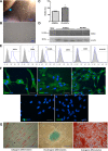
Characterization of the WJ-MSCs used. (A): Wharton’s jelly explant (blue arrow), with cells proliferating on the surface, on day 18 of incubation (magnification, ×4). (B): Cells in cultures from Wharton’s jelly (magnification, ×10). (C, D): Densitometry and immunoblotting of Klotho expression in WJ-MSCs and ADMSCs. (E): Fluorescence-activated cytometry of WJ-MSCs for CD34, CD45, HLA-DR, CD73, CD90, and CD105. (F): Immunofluorescence analysis of WJ-MSCs for CD14, CD19, CD44, CD90, and CD146. Gray line: 50 µm. (G): Analysis of the differentiation capacity of the WJ-MSCs; adipogenesis, osteogenesis, and chondrogenesis were identified by staining with Oil Red O, alizarin, and Alcian blue, respectively. Blue line: 50 µm. Abbreviations: ADMSC, adipose-derived mesenchymal stem cell; DAPI, 4′,6-diamidino-2-phenylindole; WJ-MSC, Wharton’s jelly-derived mesenchymal stem cell.
Figure 2.
Figure 2.
Survival. Survival was plotted for sham-operated rats (control group; n = 9), rats undergoing CLP and left untreated (CLP group; n = 9), and rats submitted to CLP and intraperitoneally injected, 6 hours later with 1 × 10⁶ human WJ-MSCs (CLP+WJ-MSC group; n = 9). p = .039 among groups. Abbreviations: CLP, cecal ligation and puncture; WJ-MSC, Wharton’s jelly-derived mesenchymal stem cell.
Figure 3.
Figure 3.
Inulin clearance. Renal function, as measured by inulin clearance, at 6 (A) and 24 (B) hours after CLP or sham surgery, in sham-operated rats (control group), rats submitted to CLP and left untreated (CLP group), and rats submitted to CLP and intraperitoneally injected, 6 hours later, with 1 × 10⁶ human WJ-MSCs (CLP+WJ-MSC group). ∗∗, p < .0001 versus control; ##, p < .0001 versus CLP. Abbreviations: CLP, cecal ligation and puncture; WJ-MSC, Wharton’s jelly-derived mesenchymal stem cell.
Figure 4.
Figure 4.
Macrophage infiltration and apoptosis. Representative photomicrographs (A) (magnification, ×40, blue line: 50 µm) and bar graph (B) showing CD68+ cells (red arrows) in the tubulointerstitium, at 24 hours after CLP or sham surgery, in sham-operated rats (control group; n = 8), rats submitted to CLP and left untreated (CLP group; n = 8), and rats submitted to CLP and intraperitoneally injected, 6 hours later, with 1 × 10⁶ human WJ-MSCs (CLP+WJ-MSC group; n = 8). TUNEL staining was used to detect apoptosis. Photomicrographs (C) (magnification, ×40, blue line: 50 µm) showing TUNEL+ cells (blue arrows) in the renal cortex, quantified in a bar graph (D), of control rats (n = 8), CLP rats (n = 8), and CLP+WJ-MSC rats (n = 8). Values are mean ± SEM. ∗, p < .05 versus control; #, p < .05 versus CLP. Abbreviations: CLP, cecal ligation and puncture; TUNEL, terminal deoxynucleotidyl transferase deoxyuridine triphosphate nick end labeling; WJ-MSC, Wharton’s jelly-derived mesenchymal stem cell.
Figure 5.
Figure 5.
Expression of VEGF, Klotho protein, NF-κB, eNOS, Bax protein, and Bcl-X protein. Densitometry (A–F) and immunoblotting (G) of markers of renal injury, at 24 hours after CLP or sham surgery, in sham-operated rats (control group; n = 8), rats submitted to CLP and left untreated (CLP group; n = 8), and rats submitted to CLP and intraperitoneally injected, 6 hours later, with 1 × 10⁶ human WJ-MSCs (CLP+WJ-MSC group; n = 8). Values are mean ± SEM. ∗, p < .05 versus control; ∗∗, p < .0001 versus control; #, p < .05 versus CLP; ##, p < .0001 versus CLP. Abbreviations: CLP, cecal ligation and puncture; eNOS, endothelial nitric oxide; NF-κB, nuclear factor κB; VEGF, vascular endothelial growth factor; WJ-MSC, Wharton’s jelly-derived mesenchymal stem cell.
Figure 6.
Figure 6.
Glycogen deposition in the liver. Photomicrographs of liver samples stained with periodic acid-Schiff to reveal glycogen deposits in the hepatocyte cytoplasm (A) (magnification, ×40; blue line: 50 um) and a bar graph quantifying glycogen deposition (B), at 24 hours after CLP or sham surgery, in sham-operated rats (control group; n = 8), rats submitted to CLP and left untreated (CLP group; n = 8), and rats submitted to CLP and intraperitoneally injected, 6 hours later, with 1 × 10⁶ human WJ-MSCs (CLP+WJ-MSC group; n = 8). Values are mean ± SEM. ∗∗, p < .0001 versus control; #, p < .05 versus CLP. Abbreviations: CLP, cecal ligation and puncture; WJ-MSC, Wharton’s jelly-derived mesenchymal stem cell.

References

    1. Jawad I, Lukšić I, Rafnsson SB. Assessing available information on the burden of sepsis: Global estimates of incidence, prevalence and mortality. J Glob Health. 2012;2:010404. - PMC - PubMed
    1. Dellinger RP, Levy MM, Rhodes A, et al. Surviving sepsis campaign: International guidelines for management of severe sepsis and septic shock: 2012. Crit Care Med. 2013;41:580–637. - PubMed
    1. Abraham E, Singer M. Mechanisms of sepsis-induced organ dysfunction. Crit Care Med. 2007;35:2408–2416. - PubMed
    1. Bagshaw SM, Uchino S, Bellomo R, et al. Septic acute kidney injury in critically ill patients: Clinical characteristics and outcomes. Clin J Am Soc Nephrol. 2007;2:431–439. - PubMed
    1. Zarbock A, Gomez H, Kellum JA. Sepsis-induced acute kidney injury revisited: Pathophysiology, prevention and future therapies. Curr Opin Crit Care. 2014;20:588–595. - PMC - PubMed

Publication types

MeSH terms