. 2016 Jun 9;12(6):e1005696.
doi: 10.1371/journal.ppat.1005696. eCollection 2016 Jun.

The ESX-5 System of Pathogenic Mycobacteria Is Involved In Capsule Integrity and Virulence through Its Substrate PPE10

Affiliations

The ESX-5 System of Pathogenic Mycobacteria Is Involved In Capsule Integrity and Virulence through Its Substrate PPE10

Louis S Ates et al. PLoS Pathog. .

Abstract

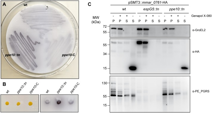
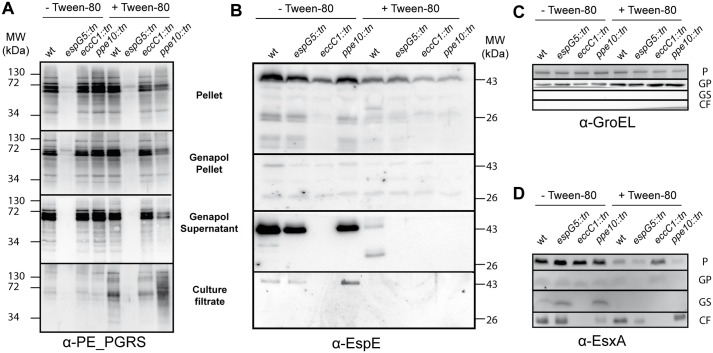
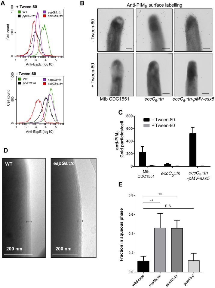
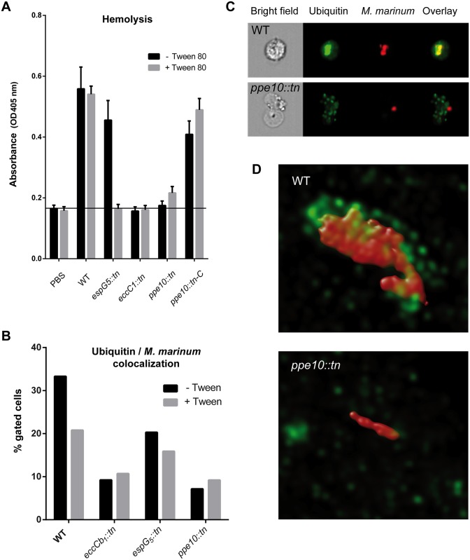
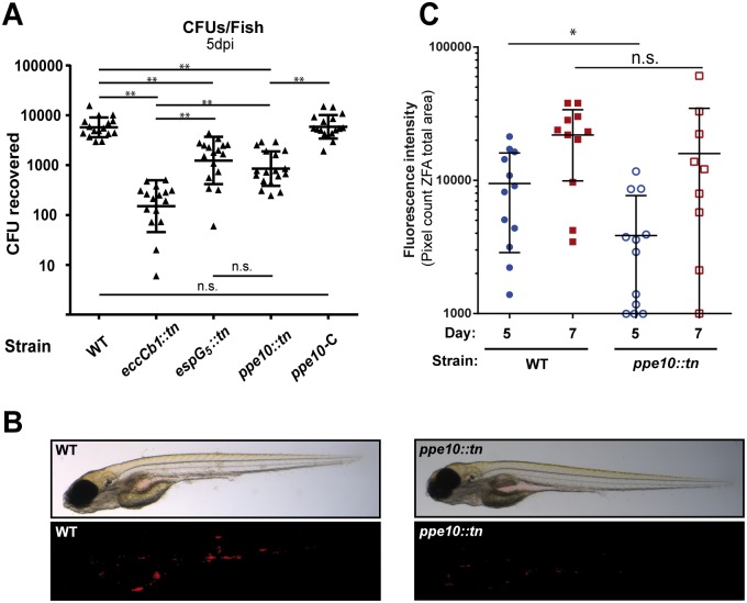
Mycobacteria produce a capsule layer, which consists of glycan-like polysaccharides and a number of specific proteins. In this study, we show that, in slow-growing mycobacteria, the type VII secretion system ESX-5 plays a major role in the integrity and stability of the capsule. We have identified PPE10 as the ESX-5 substrate responsible for this effect. Mutants in esx-5 and ppe10 both have impaired capsule integrity as well as reduced surface hydrophobicity. Electron microscopy, immunoblot and flow cytometry analyses demonstrated reduced amounts of surface localized proteins and glycolipids, and morphological differences in the capsular layer. Since capsular proteins secreted by the ESX-1 system are important virulence factors, we tested the effect of the mutations that cause capsular defects on virulence mechanisms. Both esx-5 and ppe10 mutants of Mycobacterium marinum were shown to be impaired in ESX-1-dependent hemolysis. In agreement with this, the ppe10 and esx5 mutants showed reduced recruitment of ubiquitin in early macrophage infection and intermediate attenuation in zebrafish embryos. These results provide a pivotal role for the ESX-5 secretion system and its substrate PPE10, in the capsular integrity of pathogenic mycobacteria. These findings open up new roads for research on the mycobacterial capsule and its role in virulence and immune modulation.


Conflict of interest statement

MM is employed by Eyen SE, but did not receive funding for his collaboration on this project. Eyen SE has not been financially involved in the development of this manuscript or the data and methods therein. These data do not financially benefit Eyen SE by any products, patents or other interests. This does not alter our adherence to all PLOS Pathogens policies on sharing data and materials. The other authors have declared that no competing interests exist.

Figures

Fig 1
Fig 1. Identification of ppe10::tn as a PE_PGRS supersecretor mutant and its gene product as an ESX-5 substrate.
A, B) Double filter analysis of wild-type M. marinum E11 (wt), the identified transposon mutant ppe10::tn and the complemented ppe10::tn-pSMT3::mmar_0761 (ppe10-C) strain. The double filter of a bacterial plate culture (A), or of single colonies (B) was stained with a monoclonal antibody directed against the PGRS domain. C) PPE10 is secreted in an ESX-5 dependent manner. Immunoblot with protein preparations of wild-type M. marinum E11, the ESX-5 secretion mutant espG 5::tn and the identified ppe10::tn mutant, all containing a plasmid encoding HA-tagged PPE10. The different protein preparations include cell pellets not treated with detergent Genapol X-080 (P -), cell pellets treated with Genapol X-080 (P +), Genapol X-080 supernatant fractions (S +) and culture filtrate fractions (S -). Depicted are the immunoblots stained with anti-HA antibodies or control antibodies (anti-GroEL as cell pellet marker and PE-PGRS as cell surface proteins). Full length PPE10-HA can be observed as a band with an apparent molecular weight of 58 kDa.
Fig 2
Fig 2. Both the esx-5 and ppe10 mutants release more capsular proteins when grown with Tween-80.
Wild-type M. marinum E11 (wt), and isogenic transposon mutants espG 5::tn, eccCb 1::tn and ppe10::tn were grown overnight in 7H9 medium in the presence or absence of the detergent Tween-80. Cells (P) were separated from culture filtrate (CF), after which pelleted cells were treated with Genapol X-080 to enrich for capsular proteins (Genapol supernatant (GS)). A) Immunoblot analysis with α-PE_PGRS antibody confirmed the PE_PGRS supersecretion phenotype of the ppe10::tn strain, when cells were grown in the presence of Tween-80. When grown without detergent, there is no visible phenotype of PE_PGRS secretion in the ppe10::tn strain. B) The ESX-1 substrate EspE is present in low amounts in the capsular fraction of wild-type M. marinum when grown with detergent, but this residual capsular EspE is lost in both the espG 5::tn or ppe10::tn mutant strains. C) α-GroEL was used as a loading and lysis control for all samples. D) The ESX-1 substrate EsxA could be detected in the capsular layer of cells grown without Tween-80, but seemed to be enriched in the capsule of espG 5::tn or ppe10::tn.
Fig 3
Fig 3. Surface labelling of capsular protein EspE and capsular glycans is dependent on ESX-5.
A) Parental strains of wild-type M. marinum E11, or isogenic mutant strains espG 5::tn, eccCb 1::tn and ppe10::tn expressing pSMT3::mCherry, were grown in the presence or absence of Tween-80 and labeled with the α-EspE antibody and a FITC labeled secondary antibody. Subsequently, cells were analyzed by flow cytometry analysis. High levels of surface expression of EspE could be detected in the absence of Tween-80 in both wild-type M. marinum and the espG 5::tn mutant strain, while ppe10::tn showed an intermediate EspE surface labelling. When grown in the presence of Tween-80, surface labeling of EspE was almost completely lost and reduced to similar levels of the ESX-1 mutant strain eccCb 1::tn. B) Immuno-electron microscopy analysis of M. tuberculosis CDC1551, the esx-5 mutant Mtb-eccC 5::tn and the mutant complemented with an integrative plasmid containing the complete esx-5 region; pMV-esx-5. Cells were grown in liquid culture with 0.05% Tween-80 (lower row) or without Tween-80 (upper row) and labeled with a monoclonal antibody directed against PIM6-LAM capsular glycolipids and a gold-labeled secondary antibody. C) Gold labeling of the different Mtb strains depicted in B was quantified by counting the number of gold particles per cell after growth in the presence or absence of Tween-80 (error bars indicate the standard deviation). D) The capsule morphology of wild-type M. marinum and espG 5::tn was analyzed after plunge freezing of bacteria grown in the absence of Tween-80 by cryo-electron microscopy. The black bar indicates the mycobacterial capsular layer. E) Reduced hydrophobicity of the espG 5::tn and ppe10::tn strains, measured as the OD of the aqueous phase after 1 OD of bacteria was incubated with 0.5% (v/v) Xylene in PBS for two hours. Statistical differences were calculated by GraphPad Prism software using one-way ANOVA and (Dunnett’s) multiple comparisons against a single control. ** = p<0.01, n.s = not significant.
Fig 4
Fig 4. Both the esx-5 and ppe10 mutants have reduced hemolytic activity and show reduced ubiquitin co-localization upon infection of host cells.
A) Contact-dependent hemolysis of red blood cells (RBCs) by M. marinum. M. marinum E11 wild-type cells and isogenic transposon mutants disrupted in espG 5, eccCb 1 and ppe10 and the complemented ppe10 mutant were grown in the presence or absence of Tween-80. Subsequently, washed cells were used for the hemolysis assay. B) Quantification of differentiated THP-1 cells that were infected with M. marinum. Only cells gated as positive for co-localization of the bacteria with ubiquitin were chosen for further analysis (full data and statistics in S6 Fig). The ppe10::tn mutant showed similar levels of ubiquitin co-localization as eccCb 1::tn indicating that this mutant has no cytosolic access in the early stages of infection. C) Images obtained by imaging flow cytometry of wild-type M. marinum and ppe10::tn three hours after infection of differentiated THP-1 cells. Bacteria express mCherry (Red), while ubiquitin is visualized by FK-2 antibody against poly-ubiquitin (Green). D) Super-resolution confocal microscopy images of wild-type M. marinum and ppe10::tn illustrating differential bacterial clustering and ubiquitin labeling of bacteria. Bacteria express mCherry (Red), while ubiquitin is visualized by FK-2 antibody against poly-ubiquitin (Green). A 3-dimensional view of these images can be found in supplementary S1 and S2 Movies.
Fig 5
Fig 5. The M. marinum ppe10::tn mutant is attenuated in zebrafish embryo infections.
A) M. marinum E11 wild type strain and the isogenic mutants espG 5::tn, eccCb 1::tn and ppe10::tn, as well as the complemented ppe10::tn strain (PPE10-C), were pre-cultured in liquid medium containing Tween-80. 50–100 CFUs of bacteria were injected in the bloodstream of zebrafish embryos at 28 hours post fertilization. Embryos were homogenized five days post infection and plated to establish the number of CFU per embryo. Three independent experiments of six embryos per group were performed and the data were pooled. B) Visualization of M. marinum infection of zebrafish embryos. Wild-type M. marinum or ppe10::tn containing the plasmid pSMT3::mCherry was injected in the bloodstream of zebrafish embryos as described above. After 5 days of infection the embryos were examined by brightfield (top) or fluorescence (bottom) microscopy. C) Quantification of fluorescence in infected zebrafish embryos. M. marinum wild type (closed symbols) and the ppe10::tn (open symbols) mutants expressing mCherry were injected in the bloodstream of zebrafish embryos 28 hours post fertilization. Images were acquired by fluorescence microscopy at day 5 (blue) and 7 (red) post infection and were analyzed for fluorescent intensity by dedicated software [43]. Differences on day 5 and day 7 were analyzed by GraphPad Prism software using Mann-Whitney two-tailed test. * = p<0.05, ** = p<0.01, n.s. = not significant.

References

    1. World Health Organization. Global tuberculosis report 2013. 2013.
    1. Brennan PJ, Crick DC. The cell-wall core of Mycobacterium tuberculosis in the context of drug discovery. Curr Top Med Chem. 2007;7: 475–88. Available: http://www.ncbi.nlm.nih.gov/pubmed/17346193 - PubMed
    1. Brennan PJ, Nikaido H. The envelope of mycobacteria. Annu Rev Biochem. 1995;64: 29–63. 10.1146/annurev.bi.64.070195.000333 - DOI - PubMed
    1. Hoffmann C, Leis A, Niederweis M, Plitzko JM, Engelhardt H. Disclosure of the mycobacterial outer membrane: cryo-electron tomography and vitreous sections reveal the lipid bilayer structure. Proc Natl Acad Sci U S A. 2008;105: 3963–7. 10.1073/pnas.0709530105 - DOI - PMC - PubMed
    1. Zuber B, Chami M, Houssin C, Dubochet J, Griffiths G, Daffé M. Direct visualization of the outer membrane of mycobacteria and corynebacteria in their native state. J Bacteriol. 2008;190: 5672–80. 10.1128/JB.01919-07 - DOI - PMC - PubMed

Publication types

Substances

LinkOut - more resources