

Feline Mammary Cancer
- PMID: 27281014
- PMCID: PMC7212821
- DOI: 10.1177/0300985816650243
Feline Mammary Cancer
Abstract
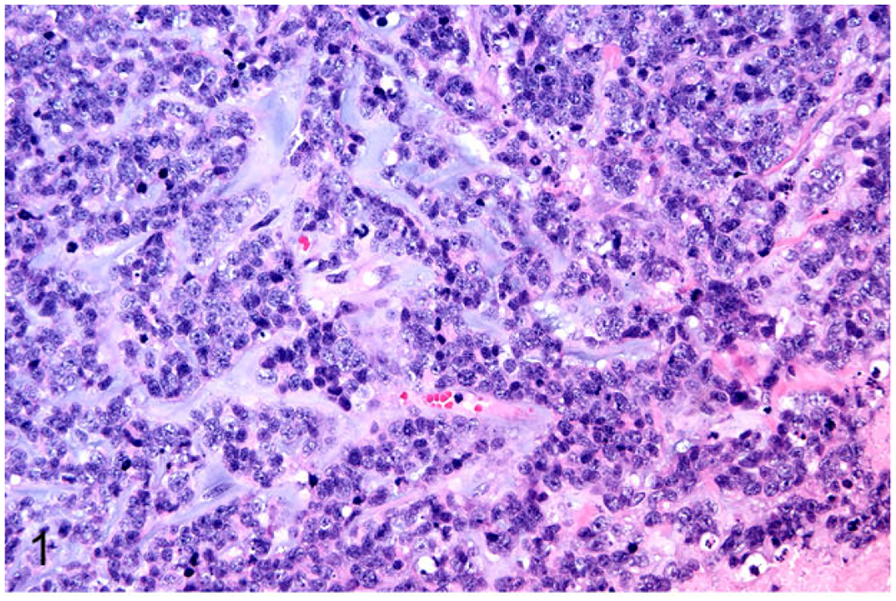
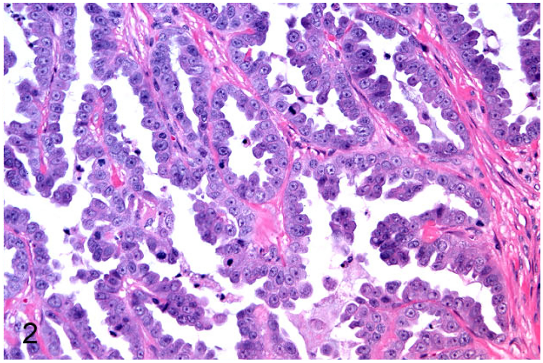
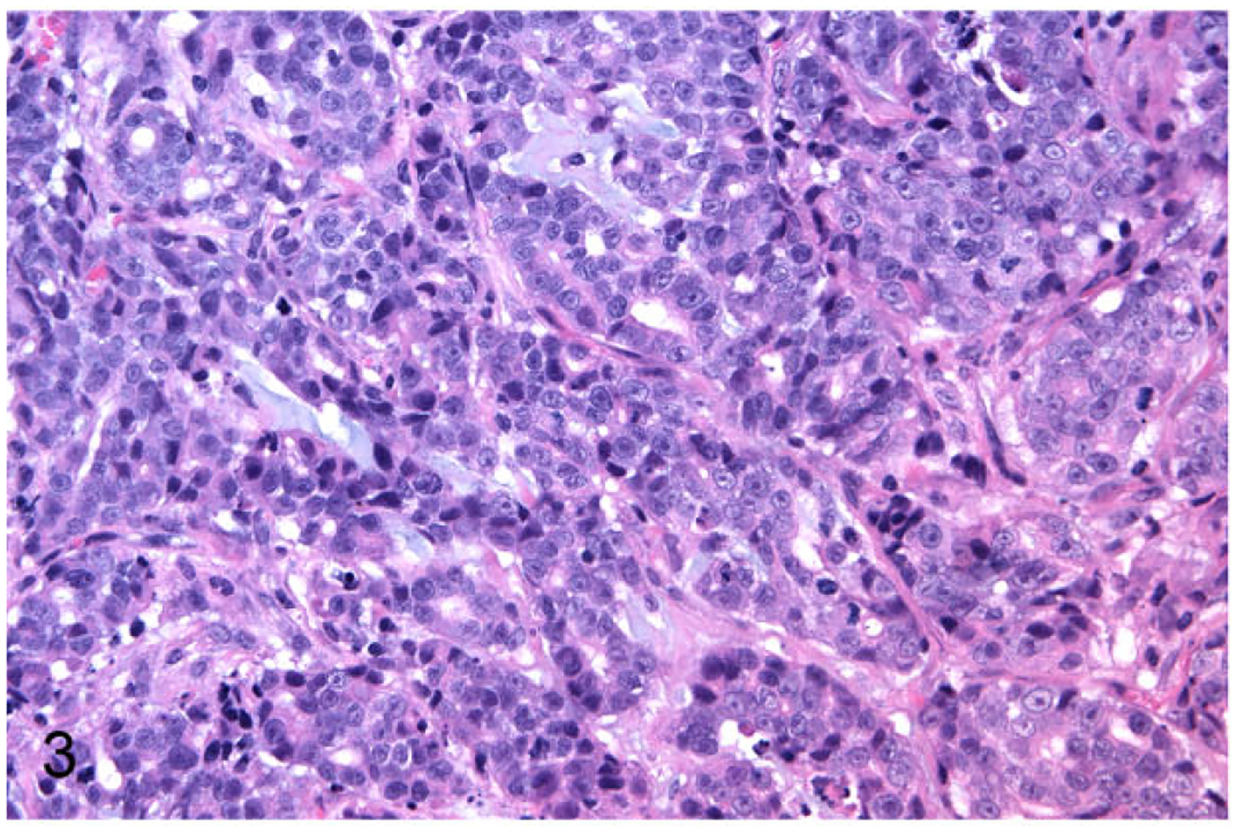
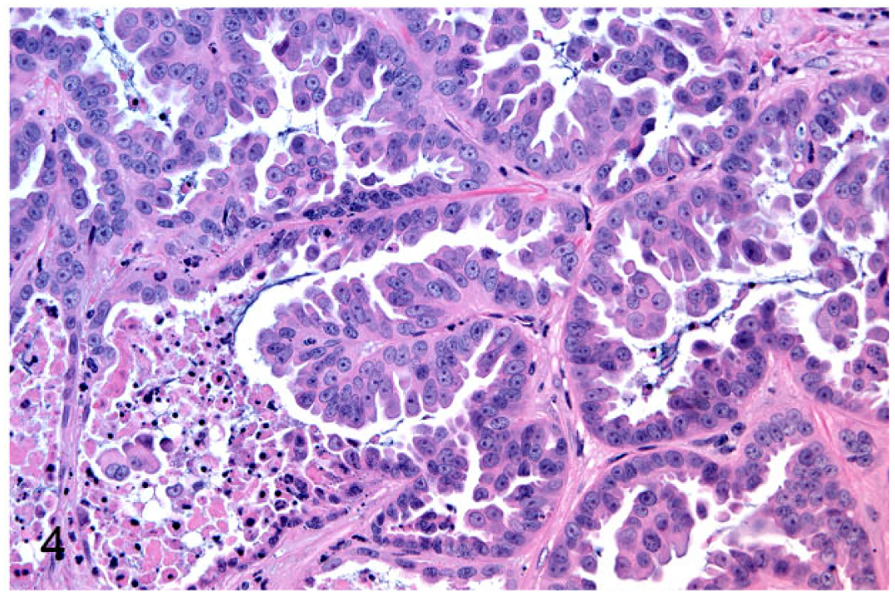
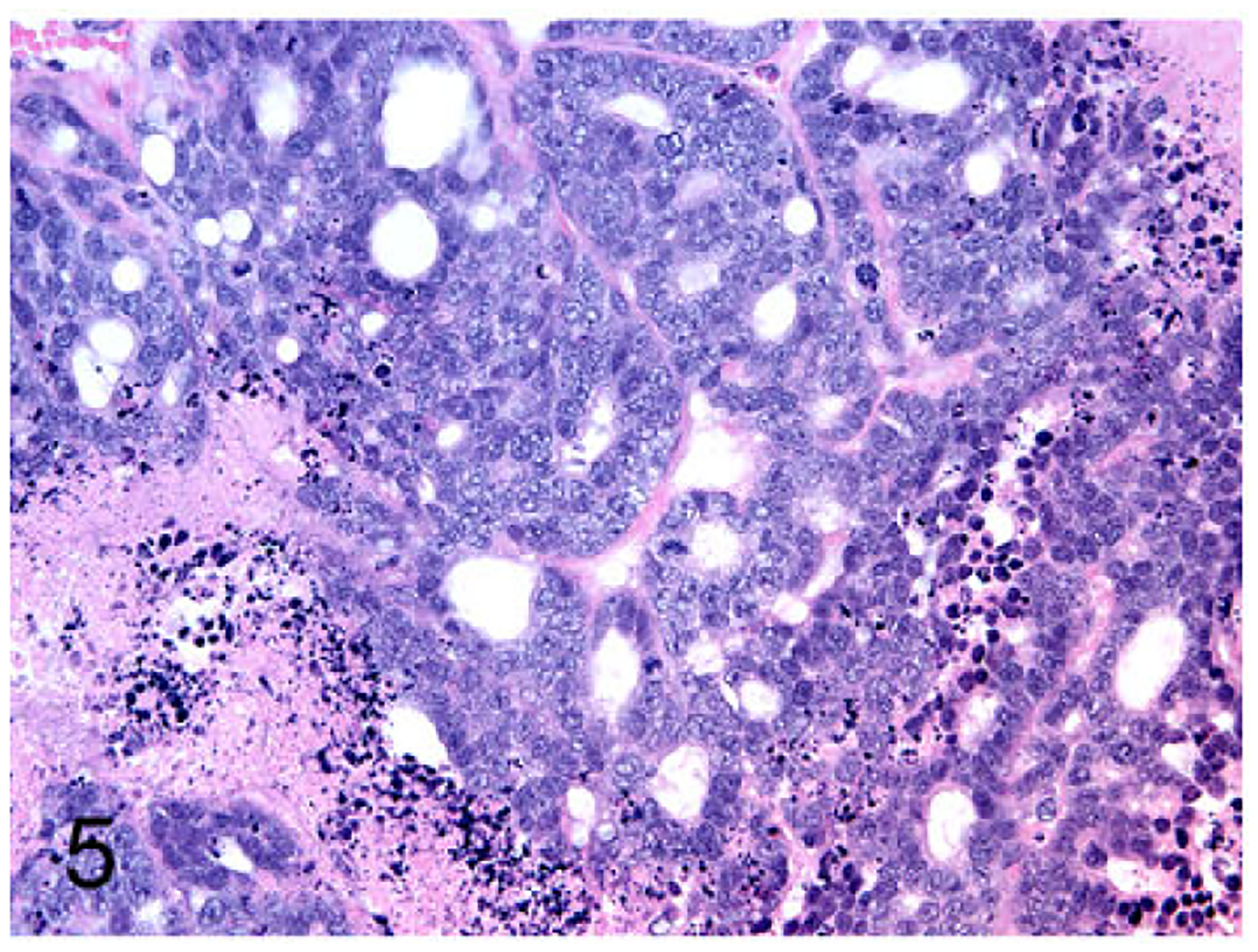
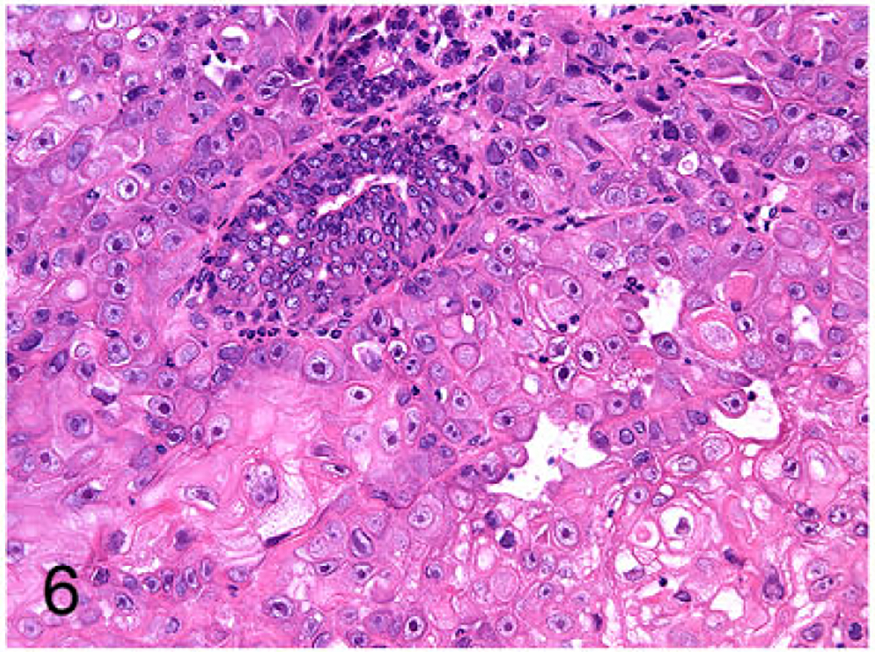
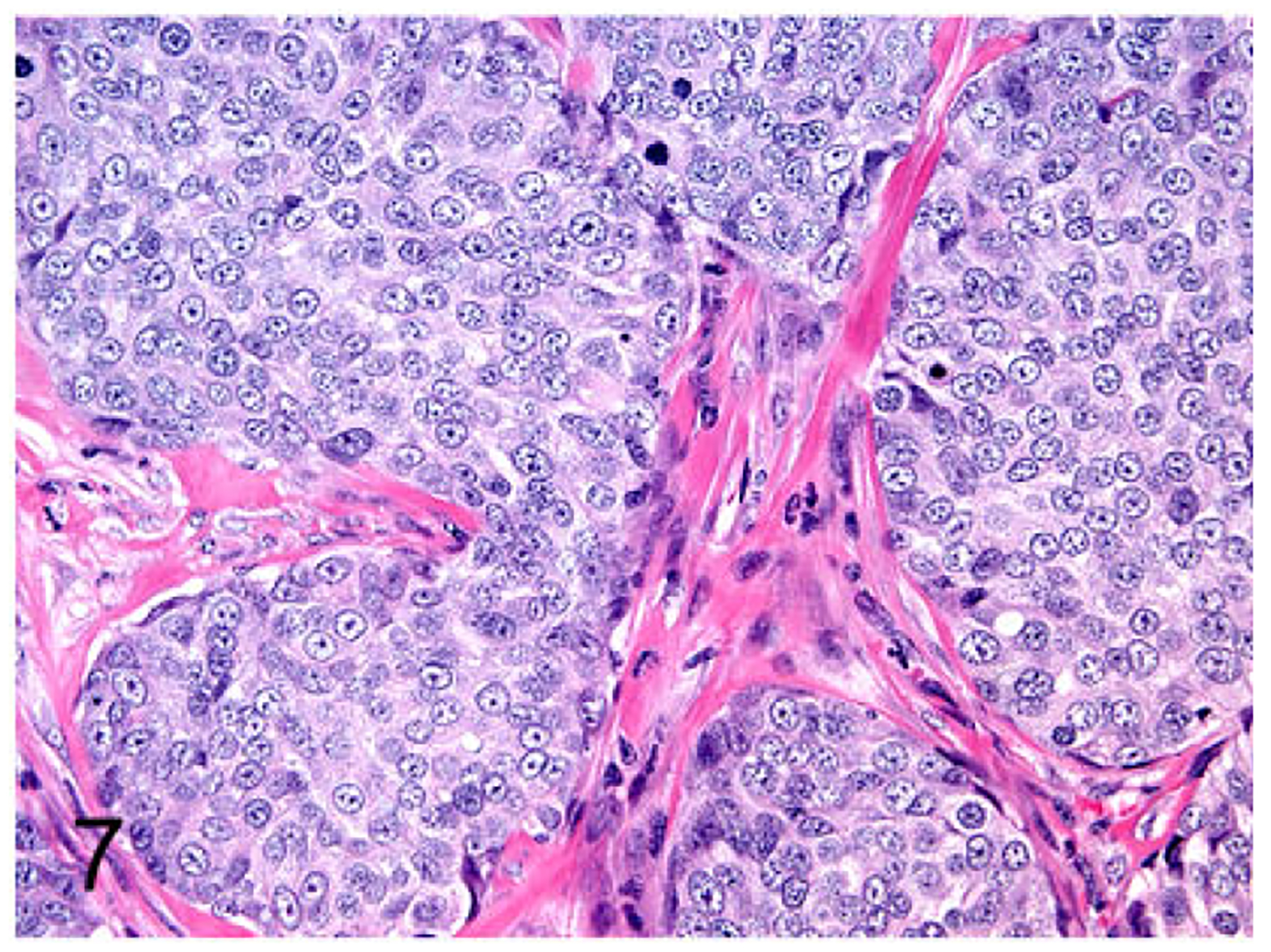
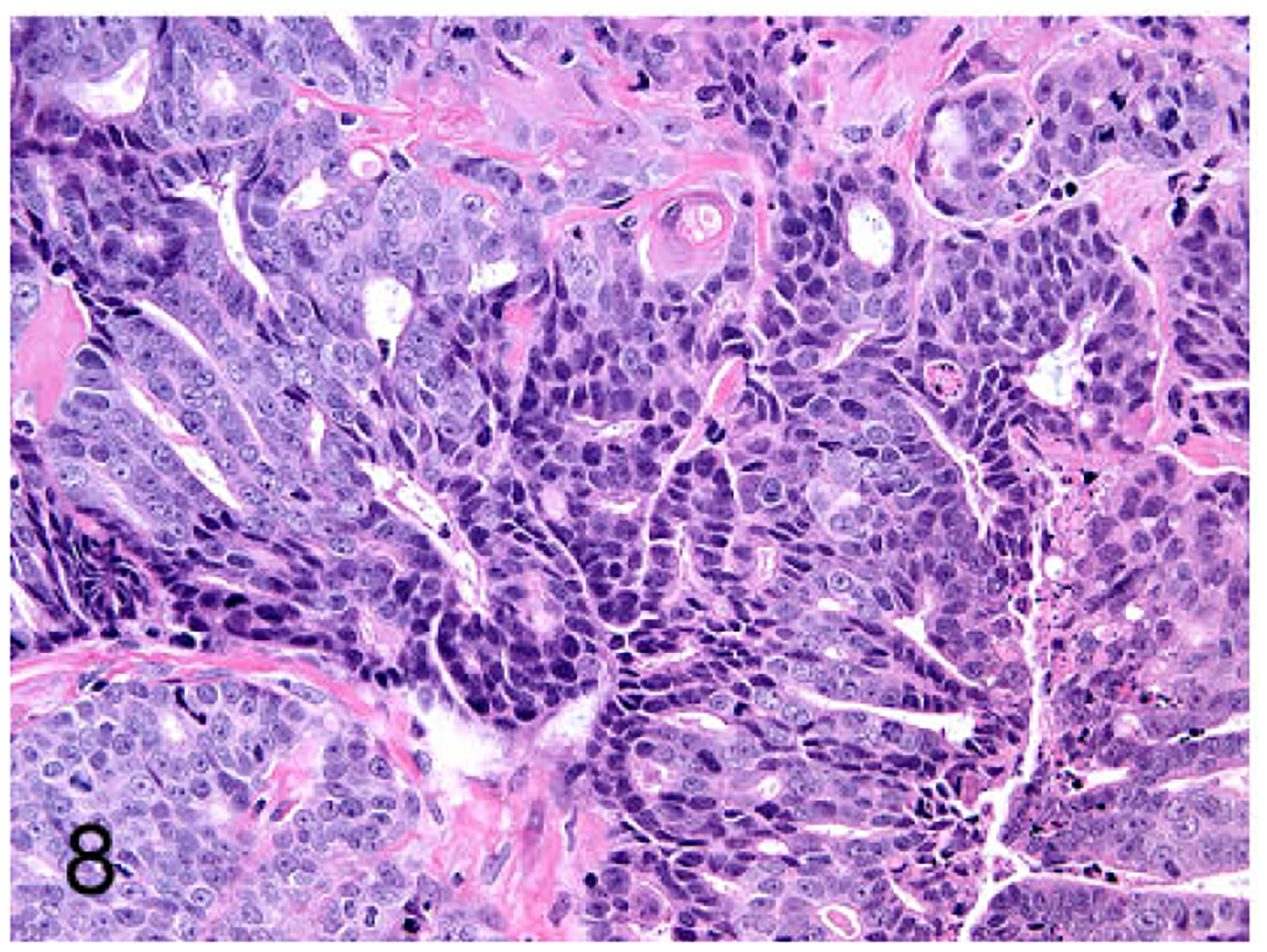
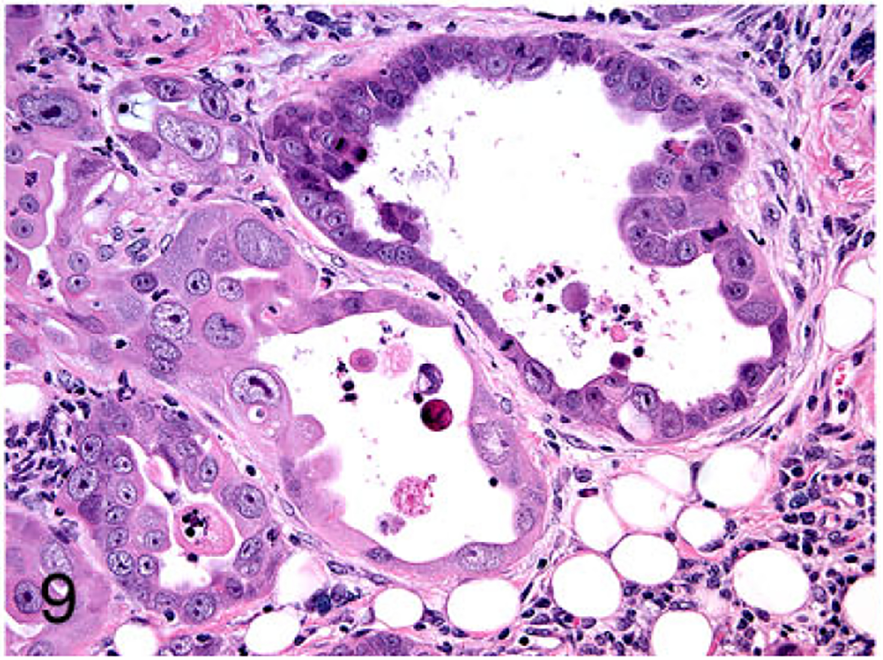
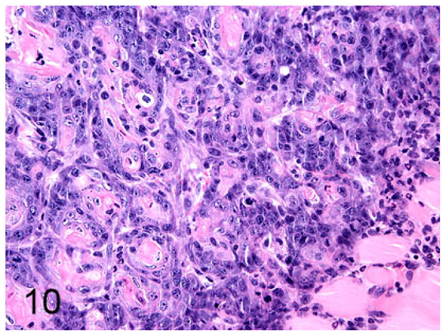
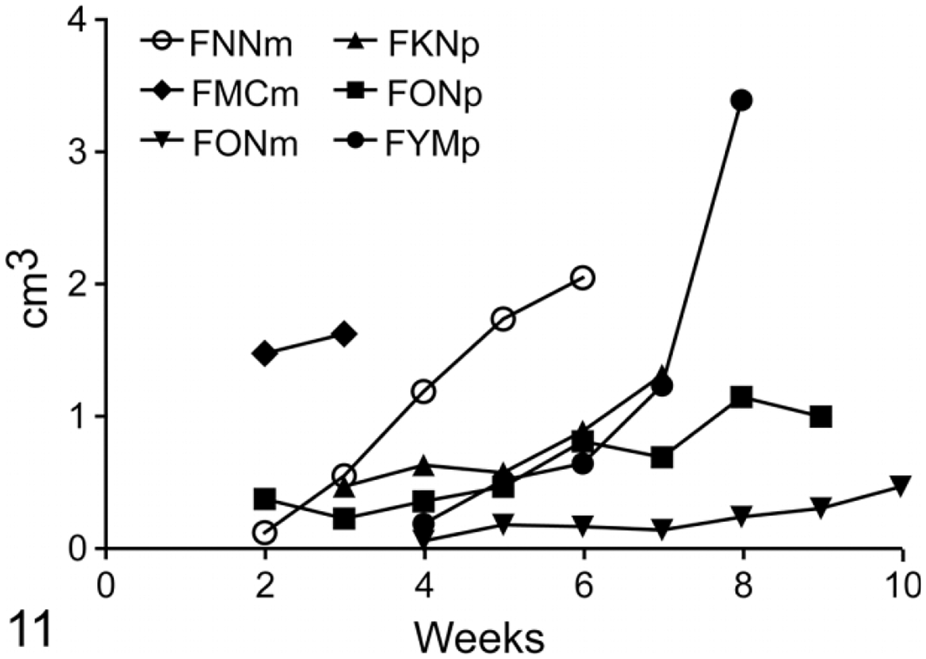
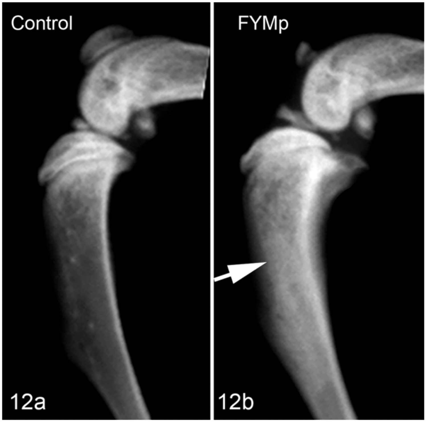
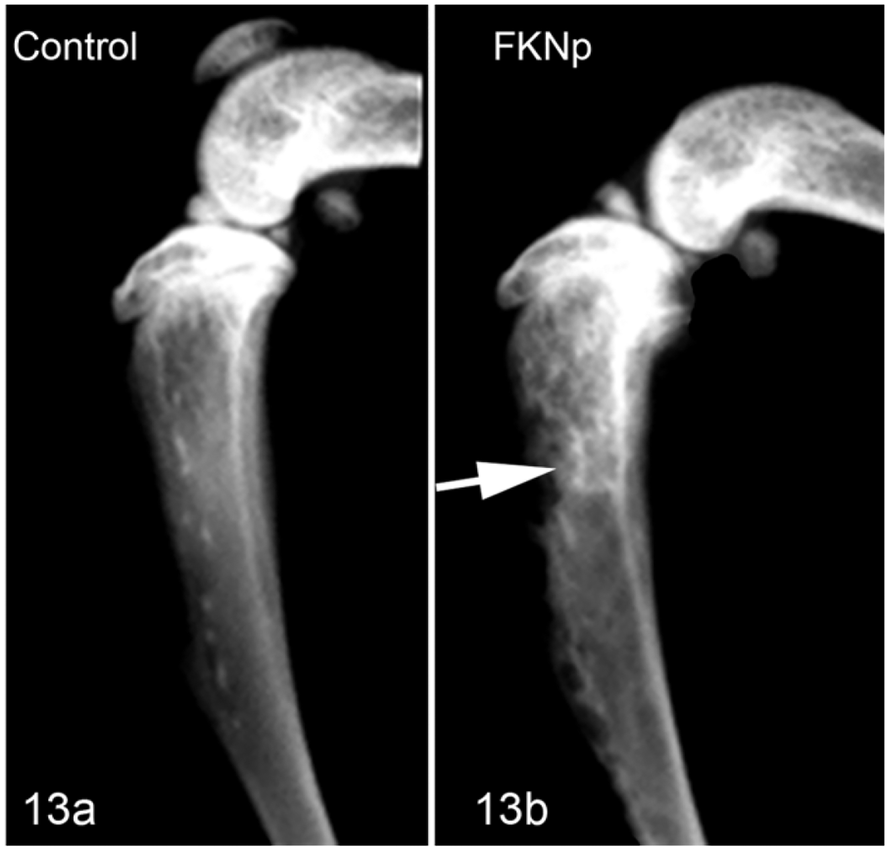
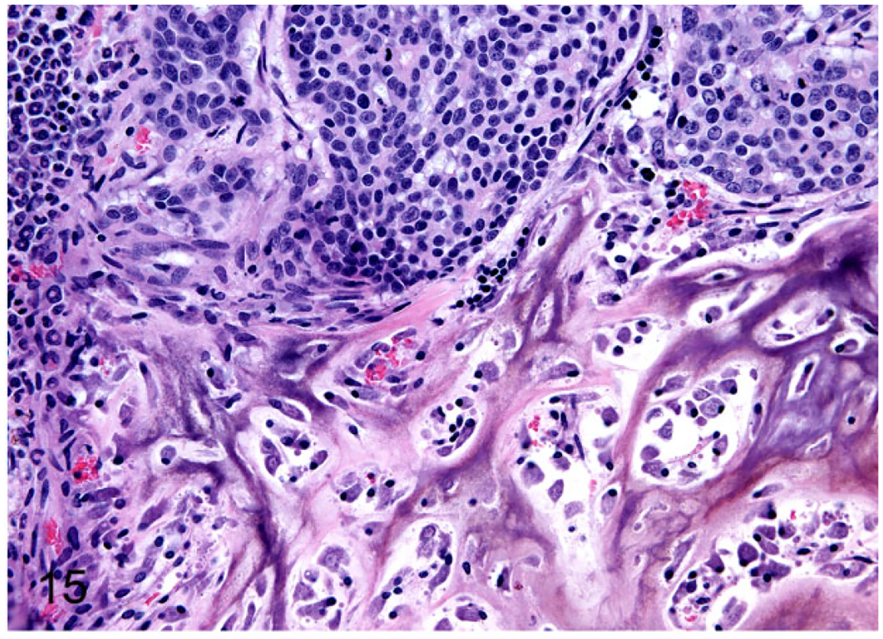
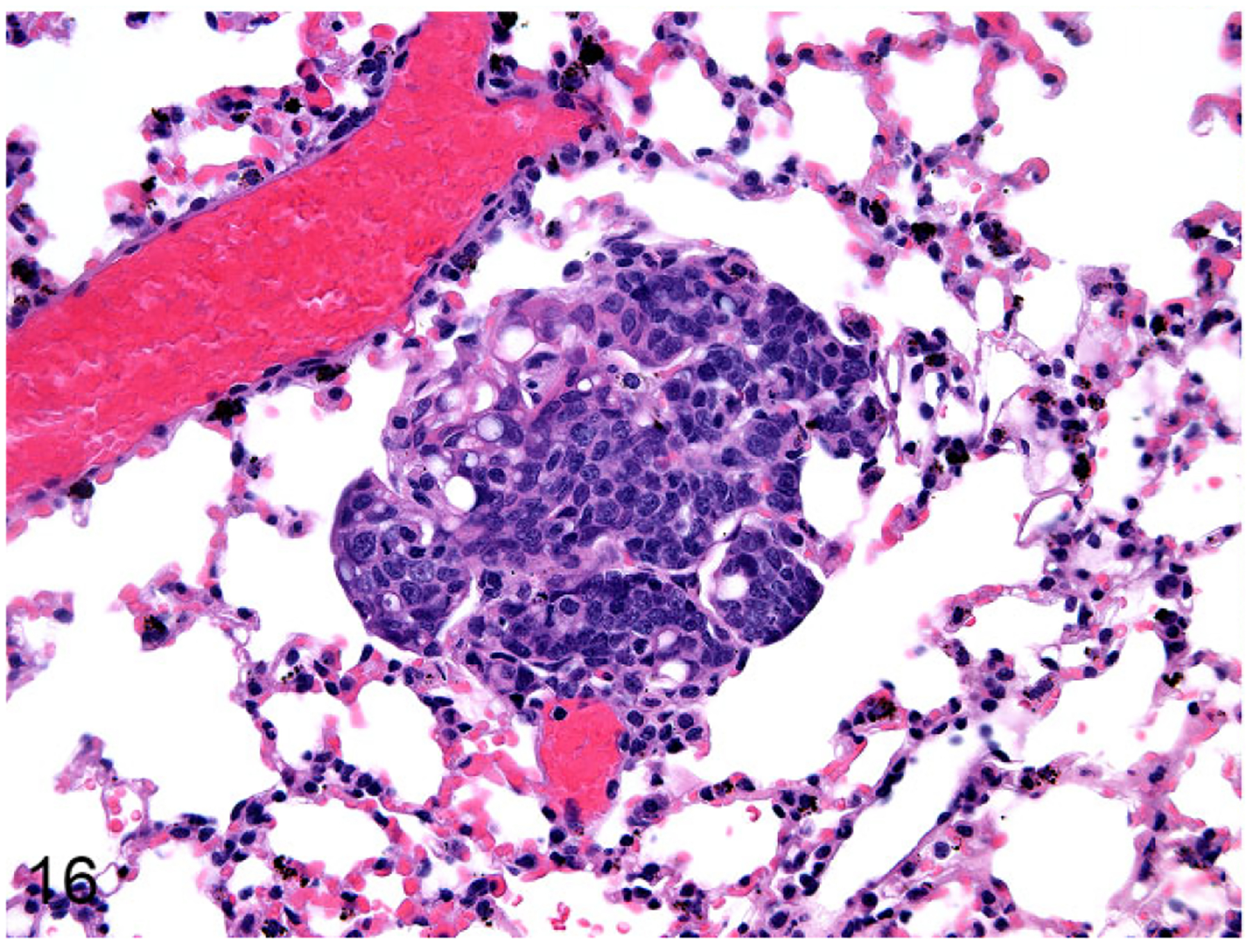
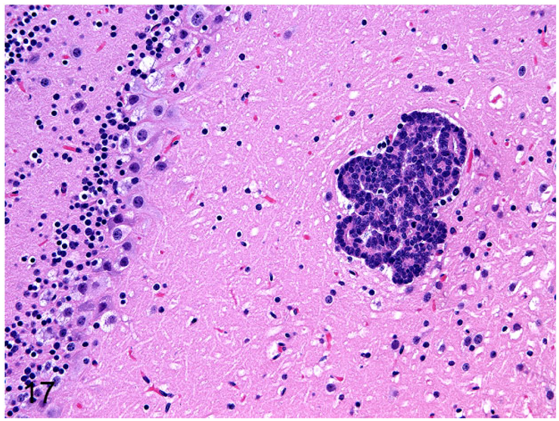
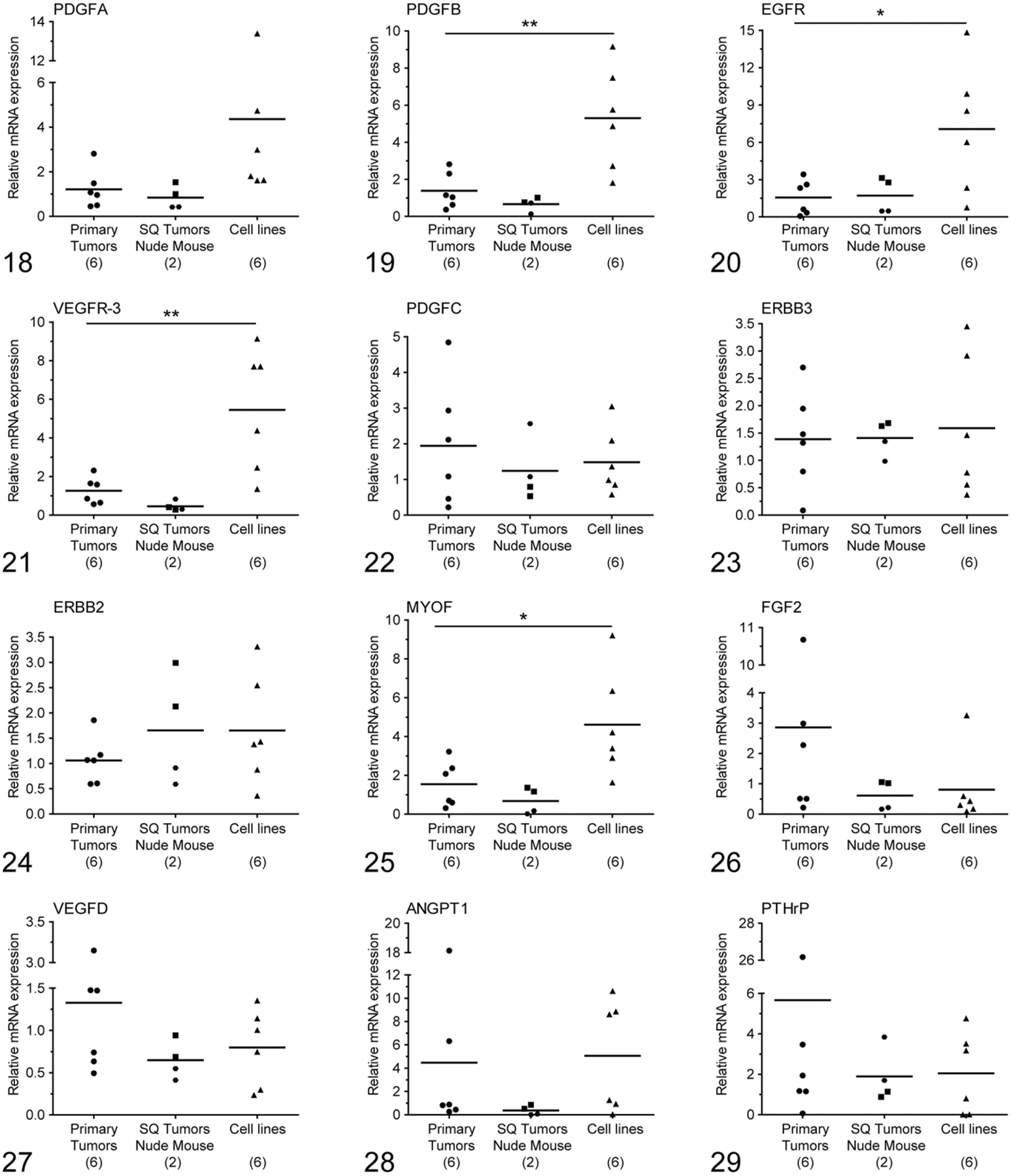
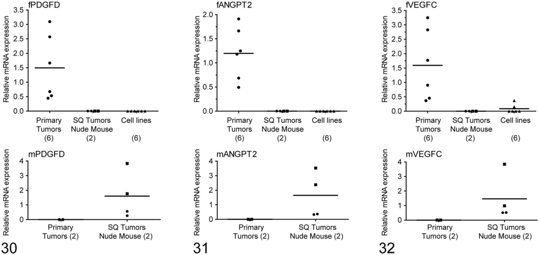
Feline mammary carcinoma (FMC) is similar to human breast cancer in the late age of onset, incidence, histopathologic features, biological behavior, and pattern of metastasis. Therefore, FMC has been proposed as a relevant model for aggressive human breast cancer. The goals of this study were to develop a nude mouse model of FMC tumor growth and metastasis and to measure the expression of genes responsible for lymphangiogenesis, angiogenesis, tumor progression, and lymph node metastasis in FMC tissues and cell lines. Two primary FMC tissues were injected subcutaneously, and 6 FMC cell lines were injected into 3 sites (subcutaneous, intratibial, and intracardiac) in nude mice. Tumors and metastases were monitored using bioluminescent imaging and characterized by gross necropsy, radiology, and histopathology. Molecular characterization of invasion and metastasis genes in FMC was conducted using quantitative real-time reverse transcription polymerase chain reaction in 6 primary FMC tissues, 2 subcutaneous FMC xenografts, and 6 FMC cell lines. The histologic appearance of the subcutaneous xenografts resembled the primary tumors. No metastasis was evident following subcutaneous injection of tumor tissues and cell lines, whereas lung, brain, liver, kidney, eye, and bone metastases were confirmed following intratibial and intracardiac injection of FMC cell lines. Finally, 15 genes were differentially expressed in the FMC tissues and cell lines. The highly expressed genes in all samples were PDGFA, PDGFB, PDGFC, FGF2, EGFR, ERBB2, ERBB3, VEGFD, VEGFR3, and MYOF. Three genes ( PDGFD, ANGPT2, and VEGFC) were confirmed to be of stromal origin. This investigation demonstrated the usefulness of nude mouse models of experimental FMC and identified molecular targets of FMC progression and metastasis.
Keywords: angiogenesis; animal model; bone; brain; cat; lung; lymphangiogenesis; mammary cancer; metastasis.
Conflict of interest statement
Declaration of Conflicting Interests
The author(s) declared no potential conflicts of interest with respect to the research, authorship, and/or publication of this article.
Figures

Similar articles
-
Bioluminescent human breast cancer cell lines that permit rapid and sensitive in vivo detection of mammary tumors and multiple metastases in immune deficient mice.Breast Cancer Res. 2005;7(4):R444-54. doi: 10.1186/bcr1026. Epub 2005 Apr 8. Breast Cancer Res. 2005. PMID: 15987449 Free PMC article.
-
Tumor microenvironment regulates metastasis and metastasis genes of mouse MMTV-PymT mammary cancer cells in vivo.Vet Pathol. 2014 Jul;51(4):868-81. doi: 10.1177/0300985813505116. Epub 2013 Oct 3. Vet Pathol. 2014. PMID: 24091811 Free PMC article.
-
Histologic evaluation of Ki-67 and cleaved caspase-3 expression in feline mammary carcinoma.J Feline Med Surg. 2017 Apr;19(4):440-445. doi: 10.1177/1098612X16634150. Epub 2016 Jul 9. J Feline Med Surg. 2017. PMID: 26917536 Free PMC article.
-
Skeletal metastasis in feline mammary carcinoma: case report and literature review.J Am Anim Hosp Assoc. 1998 Mar-Apr;34(2):103-8. doi: 10.5326/15473317-34-2-103. J Am Anim Hosp Assoc. 1998. PMID: 9507421 Review.
-
Animal models of bone metastasis.Cancer. 2003 Feb 1;97(3 Suppl):748-57. doi: 10.1002/cncr.11150. Cancer. 2003. PMID: 12548572 Review.
Cited by
-
Case report: MicroRNA-10b as a therapeutic target in feline metastatic mammary carcinoma and its implications for human clinical trials.Front Oncol. 2022 Oct 26;12:959630. doi: 10.3389/fonc.2022.959630. eCollection 2022. Front Oncol. 2022. PMID: 36387245 Free PMC article.
-
Serum PD-1/PD-L1 Levels, Tumor Expression and PD-L1 Somatic Mutations in HER2-Positive and Triple Negative Normal-Like Feline Mammary Carcinoma Subtypes.Cancers (Basel). 2020 May 28;12(6):1386. doi: 10.3390/cancers12061386. Cancers (Basel). 2020. PMID: 32481540 Free PMC article.
-
Large Animal Models of Breast Cancer.Front Oncol. 2022 Feb 4;12:788038. doi: 10.3389/fonc.2022.788038. eCollection 2022. Front Oncol. 2022. PMID: 35186735 Free PMC article. Review.
-
Trial watch: DNA-based vaccines for oncological indications.Oncoimmunology. 2017 Nov 20;6(12):e1398878. doi: 10.1080/2162402X.2017.1398878. eCollection 2017. Oncoimmunology. 2017. PMID: 29209575 Free PMC article. Review.
-
Emerging Biomarkers and Targeted Therapies in Feline Mammary Carcinoma.Vet Sci. 2021 Aug 11;8(8):164. doi: 10.3390/vetsci8080164. Vet Sci. 2021. PMID: 34437486 Free PMC article. Review.
References
-
- Alitalo K, Carmeliet P. Molecular mechanisms of lymphangiogenesis in health and disease. Cancer Cell. 2002;1(3):219–227. - PubMed
-
- Amatschek S, Koenig U, Auer H, et al. Tissue-wide expression profiling using cDNA subtraction and microarrays to identify tumor-specific genes. Cancer Res. 2004;64(3):844–856. - PubMed
-
- Bergkvist GT, Argyle DJ, Pang LY, et al. Studies on the inhibition of feline EGFR in squamous cell carcinoma: enhancement of radiosensitivity and rescue of resistance to small molecule inhibitors. Cancer Biol Ther. 2011;11(11): 927–937. - PubMed
-
- Boyde A, Maconnachie E, Reid SA, et al. Scanning electron microscopy in bone pathology: review of methods, potential and applications. Scan Electron Microsc. 1986(pt 4):1537–1554. - PubMed
Publication types
MeSH terms
Grants and funding
LinkOut - more resources
Full Text Sources
Other Literature Sources
Research Materials
Miscellaneous
