Observational Study
. 2016 Jun;95(23):e3738.
doi: 10.1097/MD.0000000000003738.

Dysfunctional phenotypes of CD4+ and CD8+ T cells are comparable in patients initiating ART during early or chronic HIV-1 infection

Affiliations
Observational Study

Dysfunctional phenotypes of CD4+ and CD8+ T cells are comparable in patients initiating ART during early or chronic HIV-1 infection

Sylvie Amu et al. Medicine (Baltimore). 2016 Jun.

Erratum in

  • Erratum: Medicine, Volume 95, Issue 23: Erratum.
    [No authors listed] [No authors listed] Medicine (Baltimore). 2016 Jul 18;95(28):e0916. doi: 10.1097/01.md.0000489580.04709.16. eCollection 2016 Jul. Medicine (Baltimore). 2016. PMID: 31265603 Free PMC article.

Abstract

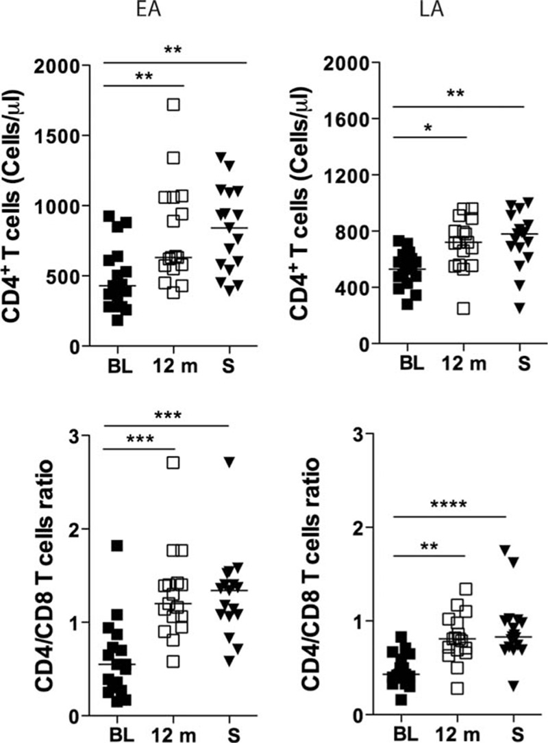
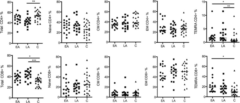
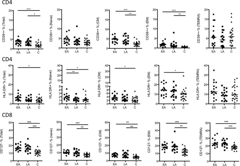
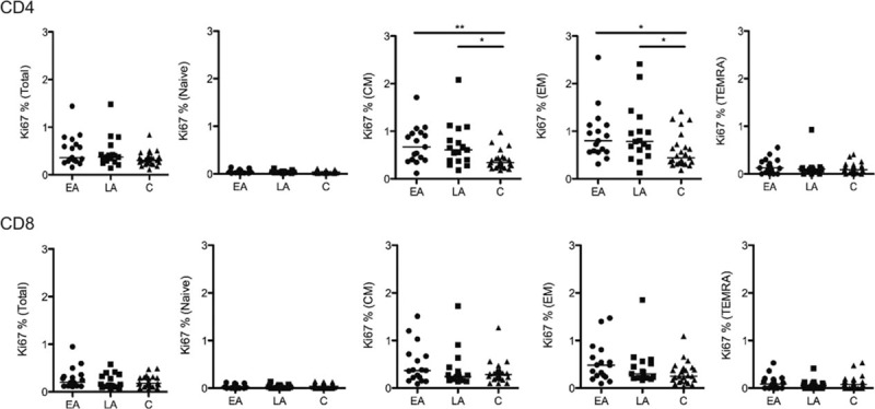
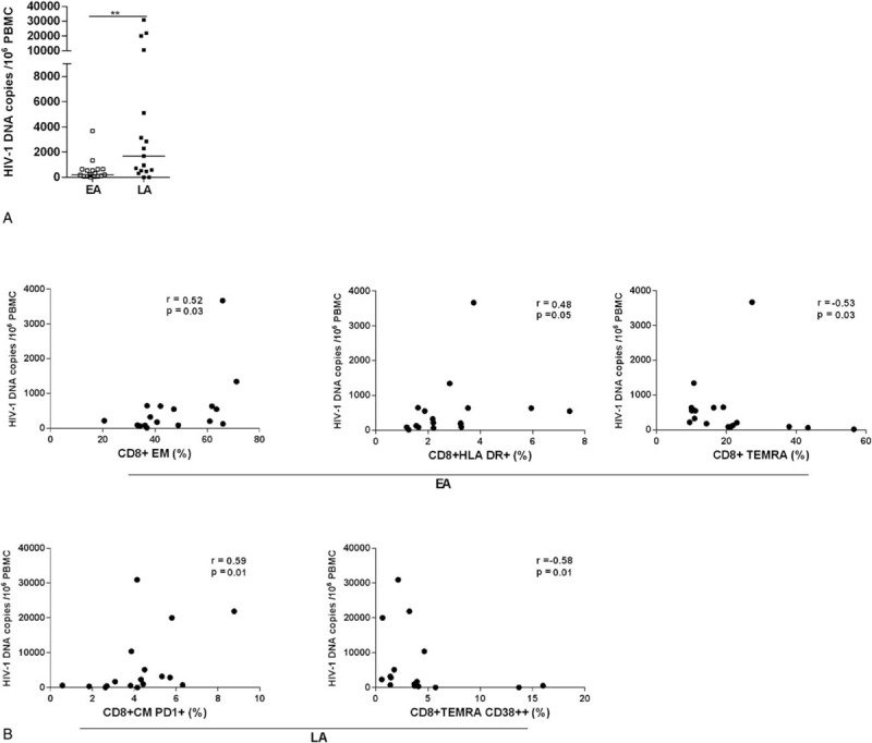
Early initiation of antiretroviral therapy (ART) is becoming a common clinical practice according to current guidelines recommending treatment to all HIV-1-infected patients. However, it is not known whether ART initiated during the early phase of infection prevents the establishment of abnormal phenotypic features previously reported in CD4+ and CD8+T cells during chronic HIV-1 infection. In this cross-sectional study, blood specimens were obtained from 17 HIV-1-infected patients who began ART treatment shortly after infection (early ART [EA]), 17 age-matched HIV-1-infected patients who started ART during chronic phase of infection (late ART [LA]), and 25 age-matched non-HIV-1-infected controls. At collection of specimens, patients in EA and LA groups had received ART for comparable periods of time. Total HIV-1 DNA was measured in white blood cells by quantitative PCR. The concentration of 9 inflammatory parameters and 1 marker of fibrosis, including sCD14 and β-2 microglobulin, was measured in plasma. Furthermore, expression of markers of abnormal immune activation (human leukocyte antigen - antigen D related [HLA-DR] and CD38), exhaustion (programmed death 1, CD28, CD57) and terminal differentiation (CD127) was measured on CD4+ and CD8+T cells. T-cell proliferation was measured through Ki67 expression. The copies of total HIV-1 DNA in blood were significantly lower (P = 0.009) in EA compared with that in LA group. Only the expression of HLA-DR on naïve CD4+ T cells distinguished EA from LA, whereas expression of 3 surface markers distinguished T-cell populations of HIV-1-infected patients from controls. These included HLA-DR distinguishing CD4+ T cells from EA compared with controls, and also CD38 and CD127 on CD4+ and CD8+ T cells, respectively, distinguishing both groups of patients from controls. The sCD14 levels were significantly higher in EA patients, and β-2 microglobulin levels were higher in LA group compared with that in controls. Our results demonstrate an equivalent abnormal expression of activation (HLA-DR and CD38 on CD4+ T cells) and terminal differentiation (CD127 on CD8+ T cells) markers in T cells from both EA and LA patients. The size of total HIV-1 DNA copies in blood of EA was lower compared with LA patients. These findings suggest that some abnormalities taking place in the T-cell compartment during primary HIV-1 infection may not be corrected by early ART.


Conflict of interest statement

The authors have no conflicts of interest to disclose.

Figures

Figure 1
Figure 1
CD4+ T-cell counts and CD4+/CD8+ ratio in the blood of individuals treated during primary (EA) and chronic (LA) phases of HIV-1 infection. CD4+ T-cell counts (cells/μL) and the ratio between CD4+ T cells and CD8+ T cells (CD4+/CD8+ ratio) were measured in the blood of subjects belonging to both EA (n = 17) and LA (n = 17) study groups. The measurements were performed before ART initiation (baseline [BL]), at 12 months (12 m) after ART and at sampling (S). The statistical difference in the values between the time points was calculated using ANOVA. Median values are shown in the figures. P < 0.05, ∗∗P < 0.01, ∗∗∗P < 0.001, and ∗∗∗∗P < 0.0001. ANOVA, analysis of variance; ART, antiretroviral therapy; EA, early ART; LA, late ART.
Figure 2
Figure 2
Frequency of CD4+ and CD8+ T cells and their subsets in PBMCs. The frequency of total CD4+ and CD8+ T cells, among CD3+ T cells, and the T-cell subsets naive, CM, EM, and TEMRA calculated out of total CD4+ or total CD8+ T cells are shown. The data are presented for all 3 groups studied: EA (n = 17), LA (n = 17), and C (n = 25). The line in the figures represents median values and the statistics were calculated using ANOVA. P < 0.05, ∗∗P < 0.01, and ∗∗∗P < 0.001. ANOVA, analysis of variance; ART, antiretroviral therapy; CM, central memory; EA, early ART; EM, effector memory; LA, late ART; TEMRA, terminally differentiated effector memory cells.
Figure 3
Figure 3
Frequency of CD38++ CD4+, HLA-DR+ CD4+, and CD127− CD8+ T-cell subsets. The frequency of CD38++ and HLA-DR+ cells among total, naive, CM, EM, and TEMRA CD4+ T cells is shown together with the frequency of CD127− total, naive, CM, EM, and TEMRA CD8+ T cells. The data are presented for the 3 groups studied: EA (n = 17), LA (n = 17), and C (n = 25). The line in the figures represents the median values and the statistics were calculated using ANOVA. P < 0.05, ∗∗P < 0.01, and ∗∗∗P < 0.001. ANOVA, analysis of variance; ART, antiretroviral therapy; CM, central memory; EA, early ART; EM, effector memory; HLA-DR, human leukocyte antigen - antigen D related; LA, late ART; TEMRA, terminally differentiated effector memory cells.
Figure 4
Figure 4
The frequency of Ki67 expressing CD4+ and CD8+ T cells. The frequency of Ki67-positive total CD4+ and CD8+ T cells and CD4+ T cells and CD8+ T-cell subsets (naive, CM, EM, and TEMRA) in the 3 groups studied are shown. The line represents median values and the differences between the groups have been calculated using ANOVA. P < 0.05 and ∗∗P < 0.01. ANOVA, analysis of variance; ART, antiretroviral therapy; CM, central memory; EM, effector memory; TEMRA, terminally differentiated effector memory cells.
Figure 5
Figure 5
Size of total HIV-1 DNA copies and its correlation to T-cell subpopulations and surface markers. Copies of HIV-1 DNA in 106 PBMCs from EA and LA patients (A). The line represents median values and the differences between the groups have been calculated using ANOVA (∗∗P < 0.01). In specimens from EA patients, the number of HIV-1 DNA copies was shown to directly correlate with the frequencies of CD8+ EM and CD8+ total HLA-DR+ T cells, whereas a negative correlation was shown between the virus reservoir and the frequency of CD8+ TEMRA T cells (B). In specimens from LA patients, the number of HIV-1 DNA copies, directly correlated to the frequency of CD8+ CM PD-1+ T cells and indirectly to the CD8+ TEMRA CD38++. Correlation coefficients and their significance were calculated using Spearman rank correlation. ANOVA, analysis of variance; ART, antiretroviral therapy; CM, central memory; EA, early ART; EM, effector memory; HLA-DR, human leukocyte antigen - antigen D related; LA, late ART; PD-1, programmed death 1; TEMRA, terminally differentiated effector memory cells.

Similar articles

Cited by

References

    1. Lundgren JD, Babiker AG, Gordin F, et al. INSIGHT START Study Group. Initiation of antiretroviral therapy in early asymptomatic HIV infection. N Engl J Med 2015; 373:795–807. - PMC - PubMed
    1. Fidler S, Porter K, Ewings F, et al. SPARTAC Trial Investigators. Short-course antiretroviral therapy in primary HIV infection. N Engl J Med 2013; 368:207–217. - PMC - PubMed
    1. Sáez-Cirión A, Bacchus C, Hocqueloux L, et al. ANRS VISCONTI Study Group. Post-treatment HIV-1 controllers with a long-term virological remission after the interruption of early initiated antiretroviral therapy ANRS VISCONTI Study. PLoS Pathog 2013; 9:e1003211. - PMC - PubMed
    1. Stöhr W, Fidler S, McClure M, et al. Duration of HIV-1 viral suppression on cessation of antiretroviral therapy in primary infection correlates with time on therapy. PLoS One 2013; 8:e78287. - PMC - PubMed
    1. Williams JP, Hurst J, Stöhr W, et al. SPARTAC Trial Investigators. HIV-1 DNA predicts disease progression and post-treatment virological control. Elife 2014; 3:e03821. - PMC - PubMed

Publication types