Review
. 2016;23(26):2908-2928.
doi: 10.2174/0929867323666160607120124.

Targeting the Microglial Signaling Pathways: New Insights in the Modulation of Neuropathic Pain

Affiliations
Review

Targeting the Microglial Signaling Pathways: New Insights in the Modulation of Neuropathic Pain

Katarzyna Popiolek-Barczyk et al. Curr Med Chem. 2016.

Abstract

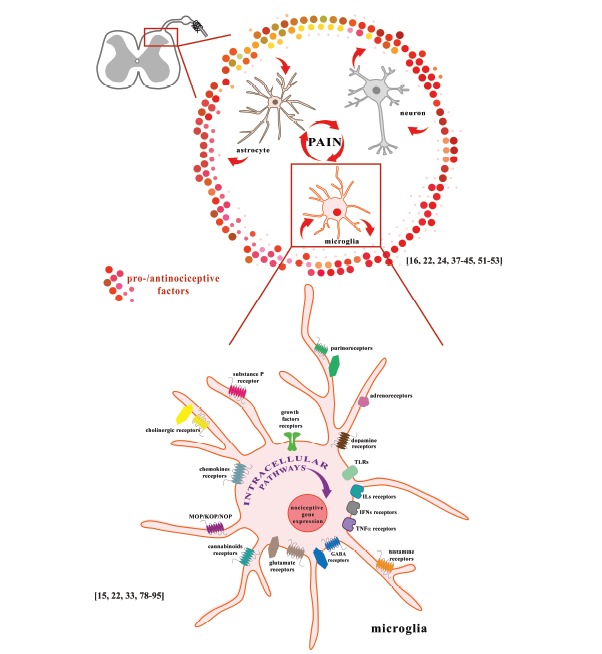
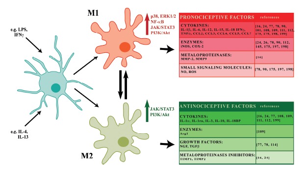
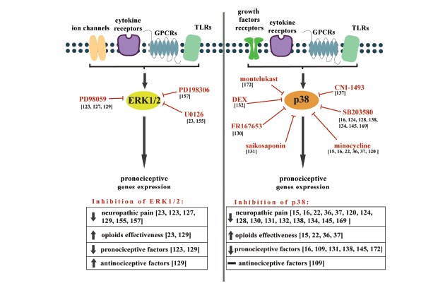
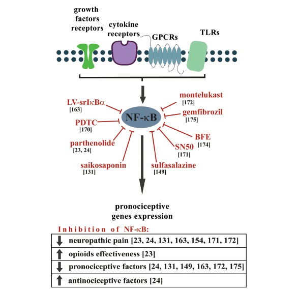
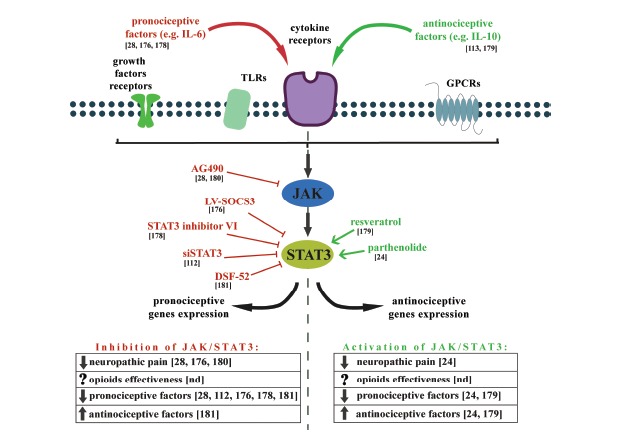
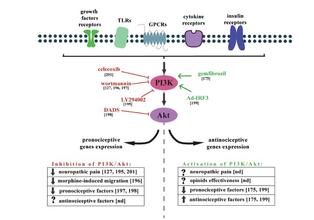
The microglia, once thought only to be supporting cells of the central nervous system (CNS), are now recognized to play essential roles in many pathologies. Many studies within the last decades indicated that the neuro-immune interaction underlies the generation and maintenance of neuropathic pain. Through a large number of receptors and signaling pathways, the microglial cells communicate with neurons, astrocytes and other cells, including those of the immune system. A disturbance or loss of CNS homeostasis causes rapid responses of the microglia, which undergo a multistage activation process. The activated microglia change their cell shapes and gene expression profiles, which induce proliferation, migration, and the production of pro- or antinociceptive factors. The cells release a large number of mediators that can act in a manner detrimental or beneficial to the surrounding cells and can indirectly alter the nociceptive signals. This review discusses the most important microglial intracellular signaling cascades (MAPKs, NF-kB, JAK/STAT, PI3K/Akt) that are essential for neuropathic pain development and maintenance. Our objective was to identify new molecular targets that may result in the development of powerful tools to control the signaling associated with neuropathic pain.


Figures

Fig. (1)
Fig. (1)
A. The interactions of neurons, astrocytes and microglia at the spinal cord level participate in the development and maintenance of neuropathic pain. B. Activation of microglial receptors leads to activation of numerous intracellular cascades. The consequence of the activation of these cells is the production of pro- and anti-nociceptive factors that are important for pain development. Microglial cells express a wide spectrum of neurotransmitter receptors: glutamate receptors (NMDA, AMPA, mGluRI, mGluRII, and mGluRIII), GABA receptors (GABAA and GABAB), cholinergic receptors (α7nAChR, M3), adrenoreceptors (α1A, α2A, β1, β2), dopamine receptors (D1-5), and purinoceptors (A1, A2A, A2B, A3, P2X1, P2X4, P2X7, P2Y2, P2Y6, P2Y12, P2Y13); receptors for hormones and modulators: histamine (H1, H2, H3, H4), opioids (MOP, KOP, NOP), cannabinoids (CB1, CB2), substance P (NK-1), neurotrophins (TrkB, TrkC), chemokines (CCR1-7, CXCR1-5, CX3CR1), interleukins (IL-1R1/IL-1R2, IL-2R, IL-4R, IL-5R, IL-6R, IL-8R, IL-9R, IL-10R, IL-12R, IL-13R, IL-15R, IL-18R), interferons (IFNγR, IFNAR type I), and TNFα (TNFR1, TNFR2); and pathogen-associated microbial patterns (TLR1-9).
Fig. (2)
Fig. (2)
Polarization of microglia. Microglial cell polarization is categorized into the classical activation state (M1), which exhibits harmful properties, and an alternative activation state (M2), which demonstrates protective and reparative functions. Depending on the environmental influences, the microglia shift their functions to maintain tissue homeostasis. Stimulation with harmful factors (e.g. LPS or IFNγ) moves the cells toward the M1 phenotype and induces the expression of proinflammatory mediators with pro-nociceptive properties (IL-1β, IL-6, IL-12, IL-15, IL-18, CCL2, CCL3, CCL4, CCL5, CCL7, IFNγ, TNFα, iNOS, COX-2, MMP-2, MMP-9, NO, ROS). However, stimulation with anti-inflammatory compounds, such as IL-4 and IL-13, promotes the M2 state, deactivates the pro-inflammatory cell phenotype, restores homeostasis and induces the increased expression of mediators with analgesic features (such as IL-1α, IL-1ra, IL-3, IL-10, IL-18BP, TIMP1, Arg1, NGF, TGFβ. Microglial cells polarization is differentially activated by intracellular signaling cascades (MAPKs, NF-κB, JAK/STAT, PI3K/Akt) that are essential for neuropathic pain development and maintenance.
Fig. (3)
Fig. (3)
The effects of ERK1/2 and p38 inhibition on neuropathic pain development and opioid effectiveness, as well as the expression of pro- and anti-nociceptive factors.
Fig. (4)
Fig. (4)
The effects of NF-κB inhibition on neuropathic pain development and opioid effectiveness, as well as the expression of pro- and anti-nociceptive factors. The NF-κB is a pleiotropic transcription factor that is activated in the responses to a wide variety of neuroinflammation-associated stimuli. The expression of the genes that are regulated by NF-κB is known to be increased following nerve injury and may contribute to the exacerbation of pain. Inhibition of NF-κB activation reduces symptoms of neuropathic pain, increases opioid effectiveness, diminishes pronociceptive and increases antinociceptive factors expression.
Fig. (5)
Fig. (5)
The effects of JAK/STAT3 modulation on neuropathic pain development and opioid effectiveness, as well as the expression of pro- and anti-nociceptive factors.
Fig. (6)
Fig. (6)
The effects of PI3K/Akt modulation on neuropathic pain development and morphine-induced microglia migration, as well as the expression of pro- and anti-nociceptive factors. The activation of PI3K/Akt pathway signaling cascade is initiated by many cytokines and growth factors as well as by insulin and LPS. This cascade modulates nociceptive information and mediates the central sensitization induced by a noxious stimuli. Inhibition of PI3K/Akt pathway activation reduces symptoms of neuropathic pain, reduces morphine-induced migration and diminishes pronociceptive factors expression. However, it was shown that also activation of PI3K/Akt pathway diminishes pronociceptive and increases antinociceptive factors.

Similar articles

Cited by

References

    1. Merskey H., Bogduk N. Classification of Chronic Pain. Seattle: IASP Press; 1994.
    1. Coyle D.E. Partial peripheral nerve injury leads to activation of astroglia and microglia which parallels the development of allodynic behavior. Glia. 1998;23:75–83. - PubMed
    1. Dworkin R.H., O’Connor A.B., Audette J., Baron R., Gourlay G.K., Haanpää M.L., Kent J.L., Krane E.J., Lebel A.A., Levy R.M., Mackey S.C., Mayer J., Miaskowski C., Raja S.N., Rice A.S., Schmader K.E., Stacey B., Stanos S., Treede R.D., Turk D.C., Walco G.A., Wells C.D. Recommendations for the pharma-cological management of neuropathic pain: an overview and literature update. Mayo Clin. Proc. 2010;85:S3–S14. - PMC - PubMed
    1. Kalso E., Allan L., Dellemijn P.L., Faura C.C., Ilias W.K., Jensen T.S., Perrot S., Plaghki L.H., Zenz M. Recommendations for using opioids in chronic non-cancer pain. Eur. J. Pain. 2003;7:381–386. - PubMed
    1. Kalso E., Edwards J.E., Moore R.A., McQuay H.J. Opioids in chronic non-cancer pain: Systematic review of efficacy and safety. Pain. 2004;112:372–380. - PubMed

MeSH terms

Substances