Review
. 2016 Oct:40:41-47.
doi: 10.1016/j.gde.2016.05.009. Epub 2016 Jun 6.

Retina regeneration in zebrafish

Affiliations
Review

Retina regeneration in zebrafish

Jin Wan et al. Curr Opin Genet Dev. 2016 Oct.

Abstract

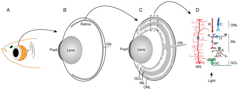
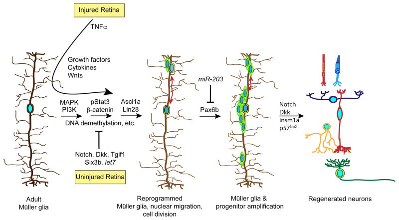
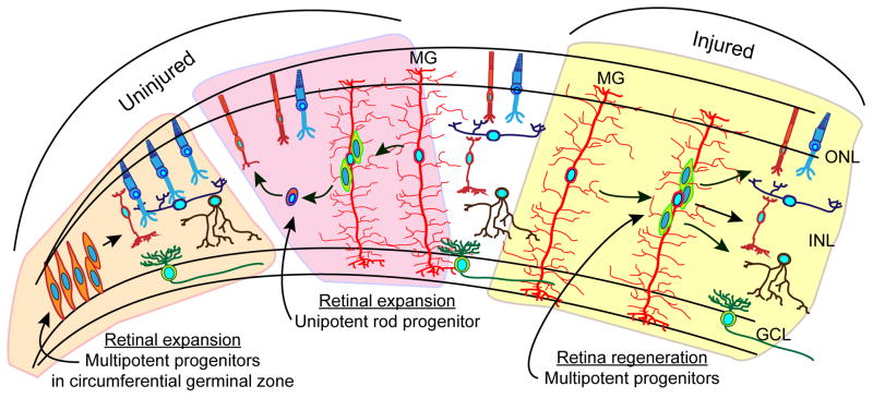
Unlike mammals, zebrafish are able to regenerate a damaged retina. Key to this regenerative response are Müller glia that respond to retinal injury by undergoing a reprogramming event that allows them to divide and generate a retinal progenitor that is multipotent and responsible for regenerating all major retinal neuron types. The fish and mammalian retina are composed of similar cell types with conserved function. Because of this it is anticipated that studies of retina regeneration in fish may suggest strategies for stimulating Müller glia reprogramming and retina regeneration in mammals. In this review we describe recent advances and future directions in retina regeneration research using zebrafish as a model system.


Figures

Figure 1
Figure 1
Retinal anatomy. Panel A: is a diagram of the fish head and eye. Panel B: is a diagram of the eye with the retina depicted as a thin piece of tissue lining the back of the eye. The optic nerve (ON) is composed of axons emanating from retinal ganglion cells. Panel C: is similar to panel B, but the retina is enlarged to show the various retinal layers. The retina is organized into three nuclear layers (outer nuclear layer [ONL], inner nuclear layer [INL] and ganglion cell layer [GCL]) that are separated by synaptic layers. Panel D: shows the neurons and glia of the retina. The ONL harbors Rod (R) and cone (C) photoreceptors. The INL harbors horizontal (H), bipolar (BP), amacrine (A) and Müller glial (MG) cells. The MG cell is unique in that it is the only retinal cell to extend processes into the ONL and GCL. The GCL harbors ganglion cells (GC) whose axons make up the optic nerve (ON).
Figure 2
Figure 2
Signaling pathways that regulate Müller glial cell reprogramming and retina regeneration. Shown are some of the pathways that have been identified to regulate Müller glia reprogramming, proliferation and progenitor differentiation. See text for details.
Figure 3
Figure 3
Retinal progenitors. Progenitors in the retinal periphery are responsible for producing all neurons except rods in the constantly expanding fish retina. Muller glia-derived unipotent rod progenitors are responsible for generating rods in the ONL of the expanding retina. Muller glial-derived multipotent progenitors are generated in the injured retina and responsible for regenerating all of the major retinal neurons.

References

    1. Goldman D. Muller glial cell reprogramming and retina regeneration. Nature reviews Neuroscience. 2014;15:431–42. - PMC - PubMed
    1. Lenkowski JR, Raymond PA. Muller glia: Stem cells for generation and regeneration of retinal neurons in teleost fish. Prog Ret Eye Res. 2014;40:94–123. - PMC - PubMed
    1. Gorsuch RA, Hyde DR. Regulation of Muller glial dependent neuronal regeneration in the damaged adult zebrafish retina. Exp Eye Res. 2014;123:131–40. - PMC - PubMed
    1. Bringmann A, et al. Cellular signaling and factors involved in Muller cell gliosis: neuroprotective and detrimental effects. Prog Retin Eye Res. 2009;28:423–51. - PubMed
    1. Reichenbach A, Bringmann A. New functions of Muller cells. Glia. 2013;61:651–78. - PubMed

Publication types

LinkOut - more resources