

Tie2 Expressing Monocytes in the Spleen of Patients with Primary Myelofibrosis
- PMID: 27281335
- PMCID: PMC4900622
- DOI: 10.1371/journal.pone.0156990
Tie2 Expressing Monocytes in the Spleen of Patients with Primary Myelofibrosis
Abstract
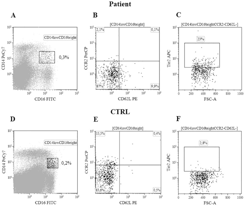
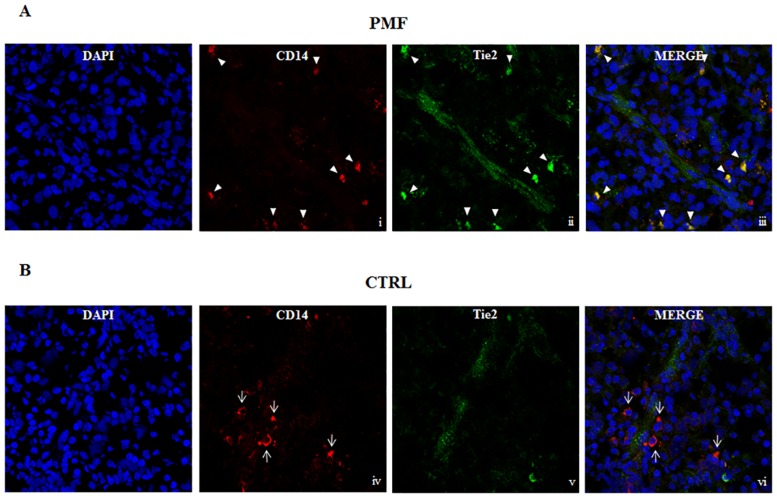
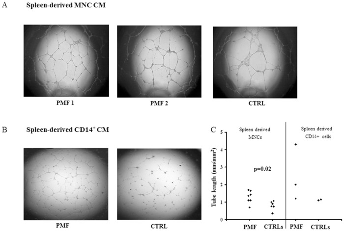
Primary myelofibrosis (PMF) is a Philadelphia-negative (Ph-) myeloproliferative disorder, showing abnormal CD34+ progenitor cell trafficking, splenomegaly, marrow fibrosis leading to extensive extramedullary haematopoiesis, and abnormal neoangiogenesis in either the bone marrow or the spleen. Monocytes expressing the angiopoietin-2 receptor (Tie2) have been shown to support abnormal angiogenic processes in solid tumors through a paracrine action that takes place in proximity to the vessels. In this study we investigated the frequency of Tie2 expressing monocytes in the spleen tissue samples of patients with PMF, and healthy subjects (CTRLs), and evaluated their possible role in favouring spleen angiogenesis. We show by confocal microscopy that in the spleen tissue of patients with PMF, but not of CTRLs, the most of the CD14+ cells are Tie2+ and are close to vessels; by flow cytometry, we found that Tie2 expressing monocytes were Tie2+CD14lowCD16brightCDL62-CCR2- (TEMs) and their frequency was higher (p = 0.008) in spleen tissue-derived mononuclear cells (MNCs) of patients with PMF than in spleen tissue-derived MNCs from CTRLs undergoing splenectomy for abdominal trauma. By in vitro angiogenesis assay we evidenced that conditioned medium of immunomagnetically selected spleen tissue derived CD14+ cells of patients with PMF induced a denser tube like net than that of CTRLs; in addition, CD14+Tie2+ cells sorted from spleen tissue derived single cell suspension of patients with PMF show a higher expression of genes involved in angiogenesis than that found in CTRLs. Our results document the enrichment of Tie2+ monocytes expressing angiogenic genes in the spleen of patients with PMF, suggesting a role for these cells in starting/maintaining the pathological angiogenesis in this organ.
Conflict of interest statement
Figures
Similar articles
-
TIE2-expressing monocytes as a diagnostic marker for hepatocellular carcinoma correlates with angiogenesis.Hepatology. 2013 Apr;57(4):1416-25. doi: 10.1002/hep.25965. Epub 2013 Feb 11. Hepatology. 2013. PMID: 22815256
-
Primary myelofibrosis marrow-derived CD14+/CD34- monocytes induce myelofibrosis-like phenotype in immunodeficient mice and give rise to megakaryocytes.PLoS One. 2019 Sep 30;14(9):e0222912. doi: 10.1371/journal.pone.0222912. eCollection 2019. PLoS One. 2019. PMID: 31569199 Free PMC article.
-
Crosstalk between TEMs and endothelial cells modulates angiogenesis and metastasis via IGF1-IGF1R signalling in epithelial ovarian cancer.Br J Cancer. 2017 Oct 24;117(9):1371-1382. doi: 10.1038/bjc.2017.297. Epub 2017 Sep 12. Br J Cancer. 2017. PMID: 28898232 Free PMC article.
-
Tie2-expressing monocytes: regulation of tumor angiogenesis and therapeutic implications.Trends Immunol. 2007 Dec;28(12):519-24. doi: 10.1016/j.it.2007.09.004. Epub 2007 Nov 5. Trends Immunol. 2007. PMID: 17981504 Review.
-
The paracrine role of Tie-2-expressing monocytes in tumor angiogenesis.Stem Cells Dev. 2009 Jun;18(5):703-6. doi: 10.1089/scd.2008.0385. Stem Cells Dev. 2009. PMID: 19186995 Review.
Cited by
-
Experimental murine acute lung injury induces increase of pulmonary TIE2-expressing macrophages.J Inflamm (Lond). 2018 Jun 14;15:12. doi: 10.1186/s12950-018-0188-5. eCollection 2018. J Inflamm (Lond). 2018. PMID: 29946226 Free PMC article.
-
Recipient Hepatic Tumor-Associated Immunologic Infiltrates Predict Outcomes After Liver Transplantation for Hepatocellular Carcinoma.Ann Transplant. 2020 Mar 13;25:e919414. doi: 10.12659/AOT.919414. Ann Transplant. 2020. PMID: 32165607 Free PMC article.
-
Monocyte Involvement in the Pathogenesis of Myeloproliferative Neoplasms.Int J Mol Sci. 2025 Jul 3;26(13):6422. doi: 10.3390/ijms26136422. Int J Mol Sci. 2025. PMID: 40650198 Free PMC article. Review.
References
-
- Barosi G, Viarengo G, Pecci A, Rosti V, Piaggio G, Marchetti M, et al. Diagnostic and clinical relevance of the number of circulating CD34(+) cells in myelofibrosis with myeloid metaplasia. Blood. 2001;98:3249–3255. - PubMed
-
- Barosi G. Myelofibrosis with myeloid metaplasia: diagnostic definition and prognostic classification for clinical studies and treatment guidelines. J Clin Oncol. 1999;17:2954–2970. - PubMed
-
- Mesa RA, Hanson CA, Rajkumar SV, Schroeder G, Tefferi A. Evaluation and clinical correlations of bone marrow angiogenesis in myelofibrosis with myeloid metaplasia. Blood. 2000;96:3374–3380. - PubMed
-
- Barosi G, Rosti V, Massa M, Viarengo GL, Pecci A, Necchi V, et al. Spleen neoangiogenesis in patients with myelofibrosis with myeloid metaplasia. Br J Haematol. 2004;124:618–625. - PubMed
Publication types
MeSH terms
Substances
LinkOut - more resources
Full Text Sources
Other Literature Sources
Research Materials
Miscellaneous
