. 2016 Jun 9;34(6):625-30.
doi: 10.1038/nbt.3606.

The US regulatory and pharmacopeia response to the global heparin contamination crisis

Affiliations

The US regulatory and pharmacopeia response to the global heparin contamination crisis

Anita Y Szajek et al. Nat Biotechnol. .

Abstract

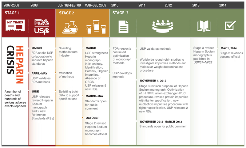
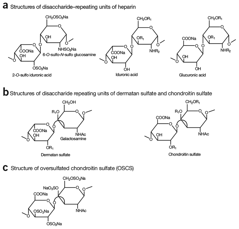
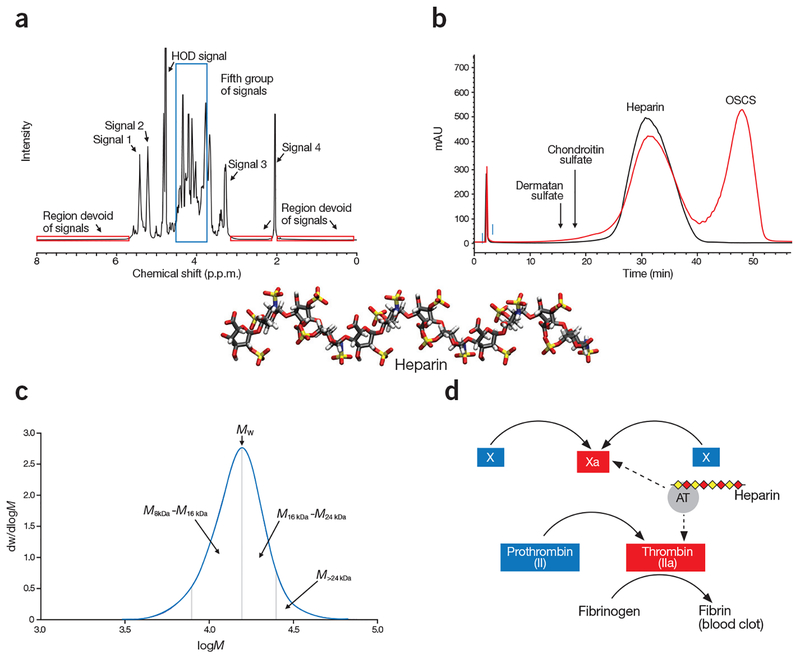
The contamination of the widely used lifesaving anticoagulant drug heparin in 2007 has drawn renewed attention to the challenges that are associated with the characterization, quality control and standardization of complex biological medicines from natural sources. Heparin is a linear, highly sulfated polysaccharide consisting of alternating glucosamine and uronic acid monosaccharide residues. Heparin has been used successfully as an injectable antithrombotic medicine since the 1930s, and its isolation from animal sources (primarily porcine intestine) as well as its manufacturing processes have not changed substantially since its introduction. The 2007 heparin contamination crisis resulted in several deaths in the United States and hundreds of adverse reactions worldwide, revealing the vulnerability of a complex global supply chain to sophisticated adulteration. This Perspective discusses how the US Food and Drug Administration (FDA), the United States Pharmacopeial Convention (USP) and international stakeholders collaborated to redefine quality expectations for heparin, thus making an important natural product better controlled and less susceptible to economically motivated adulteration.


Conflict of interest statement

COMPETING FINANCIAL INTERESTS

The authors declare competing financial interests: details are available in the online version of the paper.

Figures

Figure 1
Figure 1
Heparin crisis and resolution timeline. After the contamination crisis, the USP undertook and concluded a three-stage set of revisions to the Heparin Sodium monograph. Stage 1 involved the initial addition of identity tests and introduction of reference standards to prevent contaminated heparins from entering the US market. Stage 2 strengthened the entire monograph by including specific potency testing, additional tests and tightening existing limits for impurities. Stage 3 focused on further tightening limits for impurities and controlling heparin’s polydispersity by molecular weight analysis.
Figure 2
Figure 2
Structures of disaccharide-repeating units of glycosaminoglycans. (a) Three different disaccharide-repeating units found in heparin. The disaccharide of -IdoA2S-GlcNS6S- is most abundantly present in heparin, where IdoA2S represents 2-O-sulfo-iduronic acid; and GlcNS6S represents 6-O-sulfo-N-sulfoglucosamine. (b) The disaccharide-repeating units of dermatan sulfate and chondroitin sulfate. The disaccharide-repeating units of dermatan and chondroitin sulfates contain galactosamine. Furthermore, the uronic acid and galactosamine residues are linked through α1→3 linkages. (c) The disaccharide-repeating unit of oversulfated glycosaminoglycans, oversulfated chondroitin sulfate (OSCS), an adulterant product of chemically modified chondroitin sulfates found in contaminated heparin products. R1 = −H or −SO3Na; R2 = −H, −Ac or −SO3Na.
Figure 3
Figure 3
Orthogonal analysis of heparin. (a) Spectroscopic analysis shows a typical 1H-NMR spectrum of heparin. The specification for heparin included the identification of five groups of signals is: Signal 1 (5.42 p.p.m.) represents H1 of GlcNAc6S/GlcNS6S; Signal 2 (5.21 p.p.m.) represents H1 of IdoA2S; Signal 3 represents H2 of GlcNS (3.28 p.p.m.); and Signal 4 represents the methyl of GlcNAc (2.05 p.p.m.). The fifth group of signals (3.75–4.55 p.p.m.), corresponding to a number of heparin protons, are surrounded by a blue box. Signals 1, 2, and 4 are of comparable intensity. Signal 3 is substantially smaller than signal 2. The intensity of the fifth group of signals are 1–3 times higher than signal 2. In addition, the spectrum profile between 3.2 p.p.m. and 4.6 p.p.m., and between 4.9 p.p.m. and 5.7 p.p.m., closely matches the reference spectrum, both in intensity and signal position. The three red boxes surround regions of the spectrum where no signals should be observed. (b) Chromatographic mobility analysis showing compiled SAX–HPLC chromatograms of heparin standard (black tracing) and the mixture of HP and OSCS (red tracing). The eluted positions of other glycosaminoglycans, like dermatan sulfate and chondroitin sulfate, are indicated by arrows. (c) Molecular weight distribution analysis, shows a typical molecular weight distribution curve of heparin. In this particular determination, the weight average molecular weight (Mw) is 15,916; the proportion of material above 24 kDa (M>24 kDa) is 8.4% and the ratio of material 8–16 kDa to 16–24 kDa (M8–16 kDa/M16–24 kDa) is 1.46. dw/dlogM (y axis) represents the fraction of the material by weight. (d) Potency analysis of a partial blood coagulation cascade, demonstrating the mechanism of action of heparin. Heparin achieves its anticoagulant activity by binding to antithrombin (AT), and the heparin/AT complex inhibits the activities of factor Xa and factor IIa to prevent the formation of fibrin from fibrinogen, which is a key step in generating blood clots. The dashed arrows depict the action sites in the blood coagulation cascade by the complex of AT/heparin.

References

    1. Laremore TN, Zhang F, Dordick JS, Liu J & Linhardt RJ Recent progress and applications in glycosaminoglycan and heparin research. Curr. Opin. Chem. Biol 13, 633–640 (2009). - PMC - PubMed
    1. Lindahl U ‘Heparin’—from anticoagulant drug into the new biology. Glycoconj. J 17, 597–605 (2000). - PubMed
    1. Petitou M, Casu B & Lindahl U 1976–1983, a critical period in the history of heparin: the discovery of the antithrombin binding site. Biochimie 85, 83–89 (2003). - PubMed
    1. Barrowcliffe TW History of heparin. Handb. Exp. Pharmacol 2012, 3–22 (2012). - PubMed
    1. United States Pharmacopeia. Official monographs: Heparin Sodium (US 14) (1950)

MeSH terms