. 2016 Aug;57(8):1398-411.
doi: 10.1194/jlr.M065565. Epub 2016 Jun 8.

VNN1 promotes atherosclerosis progression in apoE-/- mice fed a high-fat/high-cholesterol diet

Affiliations

VNN1 promotes atherosclerosis progression in apoE-/- mice fed a high-fat/high-cholesterol diet

Yan-Wei Hu et al. J Lipid Res. 2016 Aug.

Abstract

Accumulated evidence shows that vanin-1 (VNN1) plays a key part in glucose metabolism. We explored the effect of VNN1 on cholesterol metabolism, inflammation, apoptosis in vitro, and progression of atherosclerotic plaques in apoE(-/-) mice. Oxidized LDL (Ox-LDL) significantly induced VNN1 expression through an ERK1/2/cyclooxygenase-2/PPARα signaling pathway. VNN1 significantly increased cellular cholesterol content and decreased apoAI and HDL-cholesterol (HDL-C)-mediated efflux by 25.16% and 23.13%, respectively, in THP-1 macrophage-derived foam cells (P < 0.05). In addition, VNN1 attenuated Ox-LDL-induced apoptosis through upregulation of expression of p53 by 59.15% and downregulation of expression of B-cell lymphoma-2 127.13% in THP-1 macrophage (P < 0.05). In vivo, apoE(-/-) mice were divided randomly into two groups and transduced with lentivirus (LV)-Mock or LV-VNN1 for 12 weeks. VNN1-treated mice showed increased liver lipid content and plasma levels of TG (124.48%), LDL-cholesterol (119.64%), TNF-α (148.74%), interleukin (IL)-1β (131.81%), and IL-6 (156.51%), whereas plasma levels of HDL-C (25.75%) were decreased significantly (P < 0.05). Consistent with these data, development of atherosclerotic lesions was increased significantly upon infection of apoE(-/-) mice with LV-VNN1. These observations suggest that VNN1 may be a promising therapeutic candidate against atherosclerosis.

Keywords: apolipoprotein E; inflammation; oxidized low density lipoprotein; vanin-1.


Figures

Fig. 1.
Fig. 1.
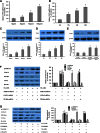
Effect and mechanism of Ox-LDL on VNN1 expression in THP-1 macrophages. A–D: Effects of Ox-LDL on VNN1 expression in THP-1 macrophages. A, C: THP-1 macrophages were treated with Ox-LDL at 0, 25, 50, and 100 μg/ml for 48 h, respectively. B, D: THP-1 macrophages were treated with 50 μg/ml of Ox-LDL for 0, 12, 24, and 48 h, respectively. Levels of VNN1 gene and protein were measured by real-time qPCR and Western blotting, respectively. E: Levels of VNN1 protein in human tissues of normal arterial intima (n = 5, 3 men; age 32.5 ± 7.8 years) and advanced atherosclerotic plaques (n = 5, 2 men; age 63.7 ± 8.9 years) were analyzed by Western blotting. F: THP-1 macrophages were transfected with control or ERK1/2 siRNA, COX-2 siRNA, or PPARα siRNA, and then incubated with 50 μg/ml Ox-LDL for 48 h. Protein expression was measured by Western blotting. G: THP-1 macrophages were transfected with control or ERK1/2 inhibitor PD98059, COX-2 inhibitor NS-398, or PPARα inhibitor GW6471, and then incubated with 50 μg/ml Ox-LDL for 48 h. Protein expression was measured by Western blotting. Data are the mean ± SD of five independent experiments, each done in triplicate. * P < 0.05 versus control.
Fig. 2.
Fig. 2.
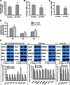
Effect of VNN1 on lipid loading, lipid content, and cholesterol efflux. THP-1 macrophages were transduced with LV-Mock or LV-VNN1. A: Then cells were incubated with 5 μg/ml of Dil-labeled Ox-LDL for 1 h and uptake of Dil-labeled Ox-LDL analyzed by flow cytometry. B, C: Cells were labeled with 0.2 μCi/ml [3H]cholesterol and cholesterol-loaded using 50 μg/ml Ox-LDL. The percentage of cholesterol efflux to apoAI (B) and HDL (C) was analyzed with a liquid scintillation counting assay. D: HPLC was done to determine the TC, free cholesterol (FC), and cholesteryl ester (CE) content in cells. E: Protein levels were measured by Western blot analyses. Data are the mean ± SD from five independent experiments, each carried out in triplicate. * P < 0.05 versus control group.
Fig. 3.
Fig. 3.
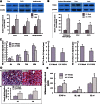
Effect and mechanism of VNN1 on apoptosis. A: THP-1 macrophages and VSMCs were transduced with LV-Mock or LV-VNN1, respectively. B: THP-1 macrophages and VSMCs were transduced with LV-Mock or LV-VNN1 and then treated with 100 μg/ml of Ox-LDL for 24 h, respectively. A, B: The proportion of apoptotic cells was assessed by flow cytometry. C, D: THP-1 macrophage were transduced with LV-Mock or LV-VNN1 and then treated with or without 100 μg/ml of Ox-LDL for 24 h as indicated. Protein levels were measured by Western blot analyses. Data are the mean ± SD from five independent experiments, each undertaken in triplicate. * P < 0.05 versus control group.
Fig. 4.
Fig. 4.
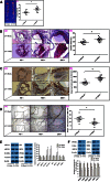
Effect of VNN1 on RCT and lipid deposition in the liver. A: C57BL/6 mice were treated with an HFD or a chow diet. VNN1 protein expression levels in various tissues were measured by Western blot. B: The expression levels of VNN1 in the LV-treated mice were measured by Western blot. C: After 12 weeks of the treatment indicated, apoE−/− mice were injected subcutaneously with [3H]cholesterol-labeled Ac-LDL-loaded BMDMs. Data are the percentage of the [3H]cholesterol tracer relative to that of total counts per minute tracer injected ± SD; n = 10, * P < 0.05 versus control group. (a) Time course of [3H]cholesterol distribution in plasma. (b) Hepatic levels of [3H]cholesterol tracer after 48 h. (c) Fecal levels of [3H]cholesterol tracer. Feces were collected continuously from 0 h to 48 h postinjection. D: (a) Liver cryosections were stained with Oil Red O. Original magnification: ×100. (b) Hepatic content of TG and TC in apoE−/− mice was assessed by enzymatic analyses. n = 10, * P < 0.05 versus control group. E: THP-1 macrophage-derived foam cells were treated with LV-Mock or LV-VNN1, respectively, and then levels of inflammatory cytokines in the medium measured with ELISA. Data are the mean ± SD from five independent experiments, each done in triplicate. * P < 0.05 versus control group.
Fig. 5.
Fig. 5.
Effect of VNN1 on the initiation and development of atherosclerosis in apoE−/− mice. A: (a) Representative staining of en face aorta with Oil Red O. (b) En face lesions were analyzed in apoE−/− mice. B: (a) Representative staining of aortic valves with Oil Red O. (b) Total lesions in the aortic valves of apoE−/− mice were analyzed. C: (a) Cryosections of aortic valves from apoE−/− mice were stained by IHC means for the macrophage marker CD68. (b) The integral optical density of CD68 in aortic-valve cryosections from apoE−/− mice was analyzed. D: (a) Representative image of aortic valves with TUNEL staining. (b) Total apoptosis in the aortic valves of apoE−/− mice was analyzed. E: Levels of protein expression in the aortic tissues of apoE−/− mice were analyzed by Western blotting. F: Levels of protein expression in peritoneal macrophages derived from apoE−/− mice were analyzed by Western blot (n = 5 mice/group). Data are the mean ± SD; n = 10. All experiments were done in triplicate, except as indicated. * P < 0.05 versus control group.
Fig. 6.
Fig. 6.
VNN1 expression and function can be regulated by Ox-LDL to promote atherosclerosis. These harmful effects are accompanied by decrease of cholesterol efflux, promotion of inflammatory molecule expression, and decrease of macrophage apoptosis, thus promoting atherosclerotic plaque formation. Ox-LDL upregulates VNN1 expression through ERK1/2/COX-2/PPARα signaling pathway. Then, VNN1 could inhibit cellular cholesterol efflux through reducing expression of ABCA1, ABCG1, SR-BI, and NPC1 by inhibiting expression of PPARγ and LXRα. In addition, VNN1 could promote inflammation through inducing expression of TNF-α, IL-1β, IL-6, ICAM-1, and VCAM-1. Moreover, VNN1 might reduce macrophage apoptosis through inhibiting P53 expression and inducing BCL-2 expression.

References

    1. Annema W., and von Eckardstein A.. 2013. High-density lipoproteins. Multifunctional but vulnerable protections from atherosclerosis. Circ. J. 77: 2432–2448. - PubMed
    1. Hu Y. W., Hu Y. R., Zhao J. Y., Li S. F., Ma X., Wu S. G., Lu J. B., Qiu Y. R., Sha Y. H., Wang Y. C., et al. . 2014. An agomir of miR-144–3p accelerates plaque formation through impairing reverse cholesterol transport and promoting pro-inflammatory cytokine production. PLoS One. 9: e94997. - PMC - PubMed
    1. Fisher E. A., Feig J. E., Hewing B., Hazen S. L., and Smith J. D.. 2012. High-density lipoprotein function, dysfunction, and reverse cholesterol transport. Arterioscler. Thromb. Vasc. Biol. 32: 2813–2820. - PMC - PubMed
    1. Hu Y. W., Ma X., Huang J. L., Mao X. R., Yang J. Y., Zhao J. Y., Li S. F., Qiu Y. R., Yang J., Zheng L., et al. . 2013. Dihydrocapsaicin attenuates plaque formation through a PPARgamma/LXRalpha pathway in apoE mice fed a high-fat/high-cholesterol diet. PLoS One. 8: e66876. - PMC - PubMed
    1. Packard R. R., and Libby P.. 2008. Inflammation in atherosclerosis: from vascular biology to biomarker discovery and risk prediction. Clin. Chem. 54: 24–38. - PubMed

Publication types

MeSH terms