. 2016 Sep 1;116(3):949-59.
doi: 10.1152/jn.00354.2016. Epub 2016 Jun 8.

Octopamine stabilizes conduction reliability of an unmyelinated axon during hypoxic stress

Affiliations

Octopamine stabilizes conduction reliability of an unmyelinated axon during hypoxic stress

T G A Money et al. J Neurophysiol. .

Abstract

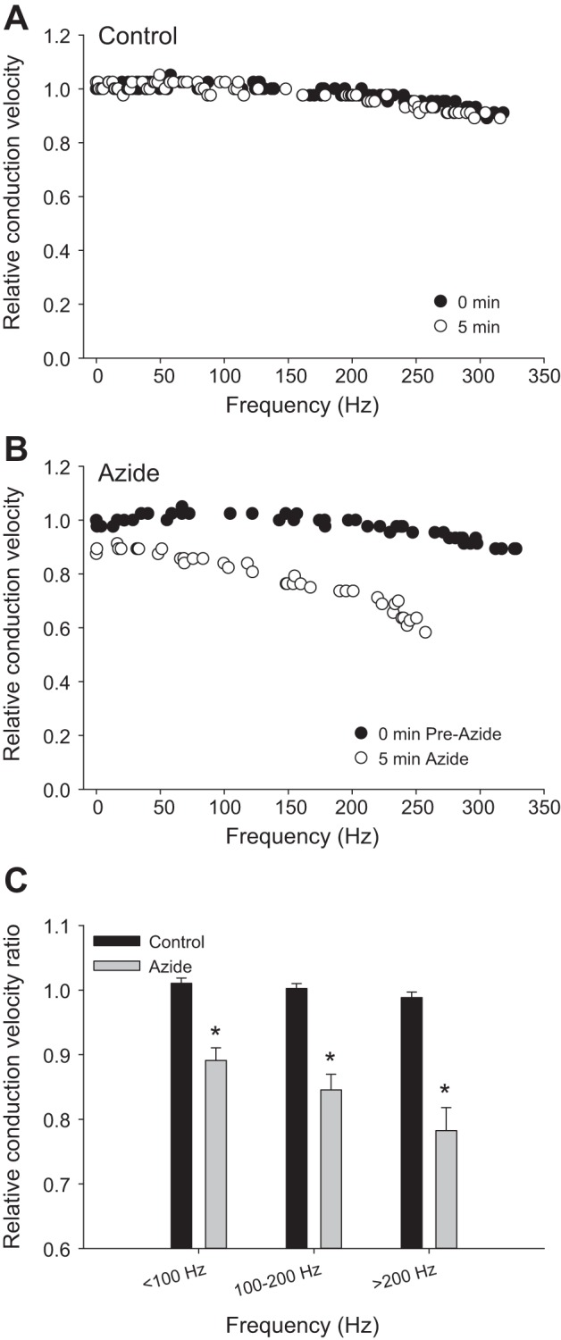
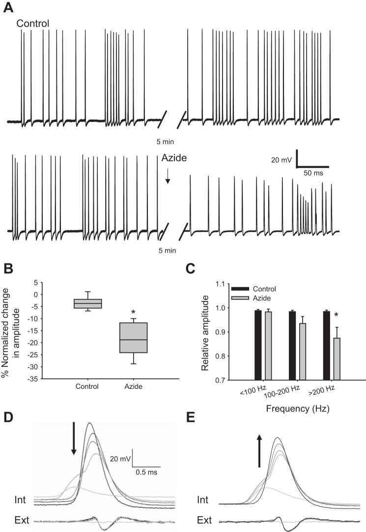
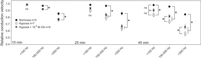
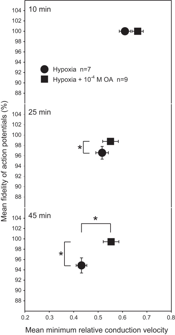
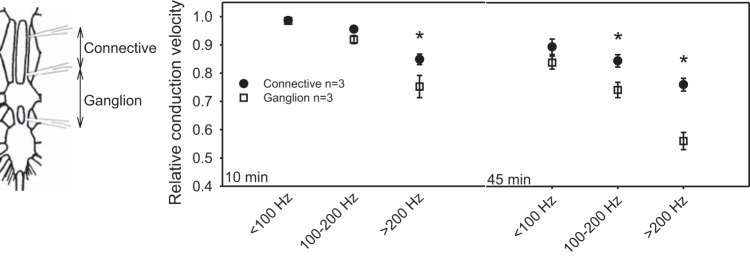
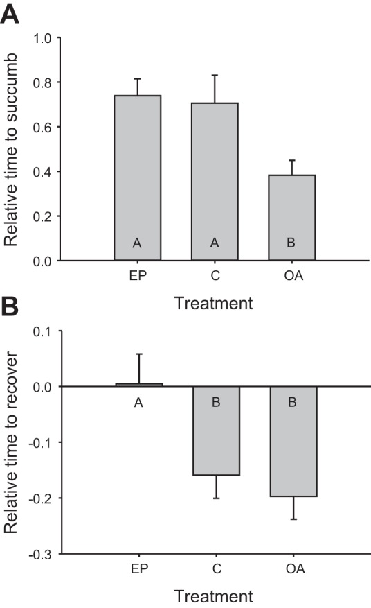
Mechanisms that could mitigate the effects of hypoxia on neuronal signaling are incompletely understood. We show that axonal performance of a locust visual interneuron varied depending on oxygen availability. To induce hypoxia, tracheae supplying the thoracic nervous system were surgically lesioned and action potentials in the axon of the descending contralateral movement detector (DCMD) neuron passing through this region were monitored extracellularly. The conduction velocity and fidelity of action potentials decreased throughout a 45-min experiment in hypoxic preparations, whereas conduction reliability remained constant when the tracheae were left intact. The reduction in conduction velocity was exacerbated for action potentials firing at high instantaneous frequencies. Bath application of octopamine mitigated the loss of conduction velocity and fidelity. Action potential conduction was more vulnerable in portions of the axon passing through the mesothoracic ganglion than in the connectives between ganglia, indicating that hypoxic modulation of the extracellular environment of the neuropil has an important role to play. In intact locusts, octopamine and its antagonist, epinastine, had effects on the entry to, and recovery from, anoxic coma consistent with octopamine increasing overall neural performance during hypoxia. These effects could have functional relevance for the animal during periods of environmental or activity-induced hypoxia.

Keywords: DCMD; action potential; conduction velocity; locust; sodium azide.


Figures

Fig. 1.
Fig. 1.
An individual response to hypoxia showing reduced conduction velocity in the DCMD axon. A: axonal action potentials recorded extracellularly in the right connective between the prothoracic (Pro) and mesothoracic (Meso) ganglia and between the Meso and metathoracic (Meta) ganglia generated in response to a 1 m/s looming object presented to the left eye. B: overlaid traces of action potentials recorded from the anterior and posterior electrodes at different times and with different instantaneous frequencies. Conduction delay between the electrodes was measured at 10 min, 25 min, and 45 min under normoxic or hypoxic conditions. C: relative conduction velocity derived from the delay measurements was stable in normoxic preparations over time and across different instantaneous firing frequencies. Hypoxic preparations, however, showed a steadily decreasing conduction velocity over time, particularly at instantaneous firing frequencies above 200 Hz.
Fig. 2.
Fig. 2.
Chemically induced metabolic stress reduces action potential conduction velocity. A: control responses to a looming object show little deterioration in action potential conduction velocity profiles over 5 min. B: 5-min treatment with 1 mM NaN3 reduced conduction velocity. C: relative conduction velocity (pre vs. 5 min) was lower during azide treatment for all action potential frequencies compared with saline control. Frequency bins: <100 Hz, 100–200 Hz, and >200 Hz. The reduction in conduction velocity was significantly greater at higher frequencies. *Significant differences between treatments, P < 0.05. nControl = 7; nAzide = 7.
Fig. 3.
Fig. 3.
Slower conduction during azide treatment is associated with a decrease in action potential amplitude. A: intracellular recording of DCMD in control saline (top) and with 1 mM azide treatment (bottom). At 5 min of exposure (bottom right), azide leads to a loss of action potential amplitude and a reduced ability to sustain high-frequency activity. B: during azide treatment, the amplitude of the lowest-frequency action potential in each response relative to pretreatment dropped significantly more than controls (Mann-Whitney rank sum test, P = 0.001). Data are plotted as median and interquartile range. C: relative amplitude ratios show that azide reduced the action potential amplitude at frequencies >200 Hz (2-way ANOVA with Holm-Sidak pairwise multiple comparisons, P < 0.001). Ratio calculated relative to the amplitude of the lowest-frequency action potential for each response. Data plotted as means ± SE. *Significant difference from control (P < 0.05). *Significant differences between treatments, P < 0.05. nControl = 6; nAzide = 7. D: raw traces of intracellular (Int) and extracellular (Ext) action potentials during azide treatment leading to conduction failure. The traces were overlaid by matching the extracellular action potential (the trace with no extracellular action potential was matched with the first sign of depolarization in the intracellular trace). During azide treatment, action potentials decrease in amplitude (arrow) until failure, evident by loss of the triphasic action potential in the extracellular trace. Note depolarization of resting membrane potential associated with shutdown of the electrogenic sodium pump. E: recovery of conduction after return to normal saline. Action potential amplitude increases (arrow) but does not return to prefailure amplitude. Note that there is no change in resting membrane potential because recovery of the sodium pump is required prior to restoration of conduction.
Fig. 4.
Fig. 4.
Octopamine (OA) stabilizes conduction velocity in the axon during hypoxia. Bath application of 10−4 M OA attenuated the hypoxia-induced loss of conduction velocity at high firing frequencies (Hypoxia vs. Hypoxia + OA). Both the Hypoxia and the Hypoxia + OA groups showed reduced conduction velocity after 45 min compared with animals with intact trachea (Normoxia). *Significance between treatments, P < 0.05. ns, No significant difference.
Fig. 5.
Fig. 5.
Octopamine reduces the incidence of conduction failure in the axon. During hypoxia, the fidelity of signaling in the DCMD decreased, with APs failing to propagate from the anterior to the posterior recording electrode. The loss was most pronounced at 45 min in hypoxic preparations, and this was reduced by 10−4 M OA bath application. *Significant differences between treatments, P < 0.05.
Fig. 6.
Fig. 6.
Reductions in conduction velocity are more pronounced during conduction through the ganglion compared with conduction within the connective. Three extracellular electrodes were used to measure conduction velocity of DCMD action potentials, both along the thoracic connective (Connective) as well as across the mesothoracic ganglion (Ganglion). During 45 min of hypoxia, conduction velocity in the axon was reduced more through the ganglion than along the connective. The difference is particularly evident at higher instantaneous firing frequencies. *Significance between treatments, P < 0.05.
Fig. 7.
Fig. 7.
Octopamine (OA) affects whole animal time to succumb and time to recover from anoxic coma. Time to succumb to anoxic coma and time to recover are expressed as a within-animal relative measure of (2nd exposure time − 1st exposure time)/1st exposure time. A: OA-treated locusts showed significantly faster times to succumb than epinastine (EP)-treated or control (C) animals. B: EP-treated animals showed significantly slower recovery times (standing upright) compared with OA-treated and control animals. Significance was assessed by a 1-way ANOVA and is indicated by letters: bars with different letters are significantly different from each other. nC = 13; nEP = 14; nOA = 12.

Similar articles

Cited by

References

    1. Armstrong GA, Rodgers CI, Money TG, Robertson RM. Suppression of spreading depression-like events in locusts by inhibition of the NO/cGMP/PKG pathway. J Neurosci 29: 8225–8235, 2009. - PMC - PubMed
    1. Armstrong GA, Shoemaker KL, Money TG, Robertson RM. Octopamine mediates thermal preconditioning of the locust ventilatory central pattern generator via a cAMP/protein kinase A signaling pathway. J Neurosci 26: 12118–12126, 2006. - PMC - PubMed
    1. Ayali A, Pener MP. Density-dependent phase polymorphism affects response to adipokinetic hormone in Locusta. Comp Biochem Physiol A 101: 549–552, 1992.
    1. Ballo AW, Nadim F, Bucher D. Dopamine modulation of Ih improves temporal fidelity of spike propagation in an unmyelinated axon. J Neurosci 32: 5106–5119, 2012. - PMC - PubMed
    1. Baylor DA, Nicholls JG. After-effects of nerve impulses on signalling in central nervous system of leech. J Physiol 203: 571–589, 1969. - PMC - PubMed

Publication types

MeSH terms

LinkOut - more resources